

Feline cataract cases require stronger etiologic thinking. Compared with dogs, cats more often have cataracts associated with uveitis, trauma, lens instability, or other ocular disease. BSAVA and Gelatt emphasize that feline cataract should not be managed by opacity alone; inflammation and systemic context matter.[1,3]
Unilateral cataract deserves caution
A unilateral cataract in a cat may reflect trauma, previous uveitis, or lens abnormality. The owner may notice only an altered pupil reflection while the cat still behaves normally with the fellow eye. Slit-lamp examination should document keratic precipitates, aqueous flare, iris color change, posterior synechiae, pupil shape, and vitreous status. These findings may predict surgical risk better than opacity density.
Why does inflammation guide the IOL decision?
If uveitis remains active, lens removal and IOL placement may increase fibrin, synechiae, and pressure instability. Inflammation should be controlled, pressure and corneal status confirmed, and surgery discussed only afterward. In cats, the question is not simply whether a model exists; it is whether the ocular environment can tolerate a long-term implant.[1,3]
How should preoperative history be taken?
Ask about trauma, chronic red eye, pain, previous medication, appetite or weight changes, and possible infectious or inflammatory disease. Examination should include pressure, slit-lamp findings, and posterior segment assessment. If the fundus cannot be seen, ultrasonography and functional assessment may be needed.
How should owners understand prognosis?
Owners can be told that the cause of cataract must be clarified first, inflammation must be quiet, and only then can lens removal and feline IOL implantation be considered. If inflammation or posterior segment disease remains unresolved, a clear-looking eye may still fail to provide stable vision.
A practical examination order for feline cataract
A useful order is to check pain and pressure first, then cornea, anterior chamber, and iris, followed by lens position and opacity, and finally fundus visibility. If anterior chamber inflammation is present, uveitis-related issues should be addressed before the clinician focuses on the lens.
Why examine a cat if the other eye seems normal?
Cats compensate well with the fellow eye, so unilateral visual loss may be noticed late. The purpose of examination is not always immediate surgery. It is to identify controllable inflammation, post-traumatic change, or pressure risk. Finding these issues early may be more valuable than waiting until both eyes are affected.
How should a feline IOL be explained?
A clear explanation is: if inflammation is quiet, the capsular bag is suitable, and the posterior segment has visual potential, a feline IOL can reduce the refractive deficit after lens removal. If the intraocular environment is unstable, the implant may add to postoperative management difficulty. This is more accurate than asking only whether a lens can be placed.
Related product note
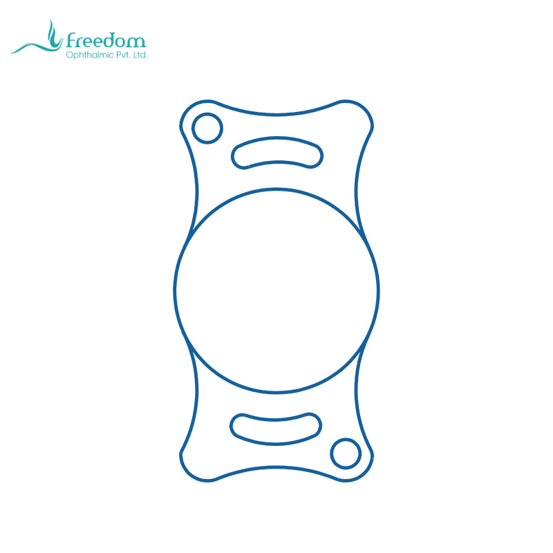
The BeneMount feline IOL is described as hydrophilic acrylic, aspheric, and foldable, with +53.50D power, a 7.00 mm optic, and 12.00/13.00/14.00 mm overall lengths. It can be product information for suitable cases, but it does not replace etiologic and inflammatory assessment.[10]