

Low IOP Does Not Mean the Eye Is Fine
Veterinarians are naturally alert to high IOP because it suggests glaucoma. In a red, painful, miotic eye, however, low IOP may be just as useful. Slatter describes hypotony, miosis, rubeosis iridis, aqueous flare, and keratic precipitates as findings that should be actively sought when anterior uveitis is suspected [2].
During anterior uveitis, inflammation affects the ciliary body and reduces aqueous humor production. Breakdown of the blood-aqueous barrier allows protein, cells, and inflammatory mediators to enter the anterior chamber. The eye may be red and painful while the pressure is lower than normal or lower than the fellow eye.
Put Low IOP into the Whole Eye Examination
Low IOP should be interpreted with the rest of the eye. Evaluate pupil size, iris color, anterior chamber clarity, aqueous flare, keratic precipitates, corneal edema, lens status, and whether the fundus can be examined. Mature or hypermature cataract, lens capsule rupture, trauma, infectious disease, and immune-mediated disease may all be associated with uveitis.
Do not reach for topical corticosteroids before corneal ulceration has been excluded. A positive fluorescein stain changes the plan. Gelatt and BSAVA place anterior uveitis at the intersection of local ocular disease and systemic workup because it may be primary, secondary to another ocular lesion, or a manifestation of systemic disease [1,3].
Follow the Trend, Not Just the First Number
The first low IOP value is a baseline. If pain improves, aqueous flare decreases, the pupil becomes more comfortable, and IOP moves closer to the fellow eye, treatment is probably controlling inflammation. If redness persists, the anterior chamber remains cloudy, or IOP begins to rise, secondary glaucoma must be considered.
BSAVA emphasizes that uveitis may later become complicated by glaucoma and that routine IOP measurement helps prevent this change from being missed [3]. Recheck records should therefore include OD/OS values and anterior chamber findings, not just “better.”
Know the Risk Boundaries
Hypotony may also occur with globe perforation, severe trauma, early postoperative leakage, impaired ciliary body function, or phthisis. Deep corneal ulceration, a suspicious Seidel test, shallow anterior chamber, iris prolapse, or recent intraocular surgery should make handling and measurement more cautious.
A single number should not override the clinical picture. If the reading is very low but the eye does not look uveitic, repeat the measurement and check device setup, probe position, corneal status, and restraint. If the reading is low and the eye is painful, earlier referral is often appropriate.
Record What Will Matter at Recheck
For low-IOP cases, document IOP in both eyes, flare, pupil status, fluorescein result, lens opacity or instability, drugs prescribed, recheck interval, and referral advice. This helps the whole team avoid treating recurrent “red eye” blindly.
The useful information is not the number alone. It is the relationship between the number, the inflammatory signs, the medication response, and the next decision.
Related Product Information
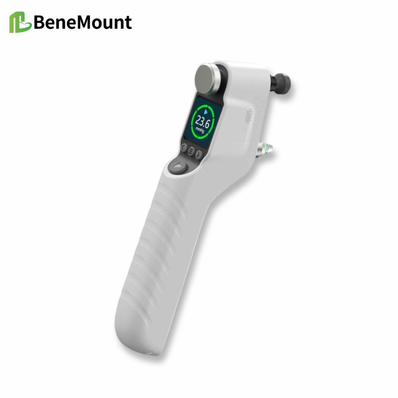
This week’s product materials include the iFalcon V100 Veterinary Tonometer and the Hawkeye V700 veterinary tonometer manuals, quick guides, and brochures. The manuals describe a rebound tonometer for animal IOP measurement using disposable TVP40 probes. During measurement, the probe should be aligned perpendicularly with the central cornea, with the probe tip about 5-7 mm from the corneal surface.
The stated animal modes include feline, canine, equine, lapine, rat, mouse, and primate. The listed measurement range is 5.0-70.0 mmHg, display range 0-99.9 mmHg, and display resolution 0.1 mmHg. For feline, canine, equine, lapine, and primate modes, the stated accuracy is ±1.2 mmHg when IOP is below 20 mmHg and ±2.2 mmHg when IOP is above 20 mmHg; for rats and mice, it is ±10%.
The device forms a final result after six successful measurements, using the best four readings and discarding two outliers according to the manual. Clinically, the number should still be interpreted with the case context: signs, corneal status, anterior chamber findings, medications, restraint, species mode, and repeatability.
References
Gelatt KN, Gilger BC, Kern TJ, et al. Veterinary Ophthalmology. 6th ed. Wiley-Blackwell; 2021. Chapters on ophthalmic examination, tonometry, canine glaucoma, and anterior uveal disease.
Maggs DJ, Miller PE, Ofri R. Slatter's Fundamentals of Veterinary Ophthalmology. 4th ed. Saunders Elsevier; 2008. Chapters on ophthalmic examination, tonometry, uveitis, and glaucoma.
Gould D, McLellan G, eds. BSAVA Manual of Canine and Feline Ophthalmology. 3rd ed. BSAVA; 2014. Chapters on ocular examination, uveal tract disease, glaucoma, and problem-oriented presentations.
Gelatt KN, Gilger BC, Kern TJ, et al. Veterinary Ophthalmology. 6th ed. Wiley-Blackwell; 2021. Chapter 21, diseases of the canine anterior uvea.