

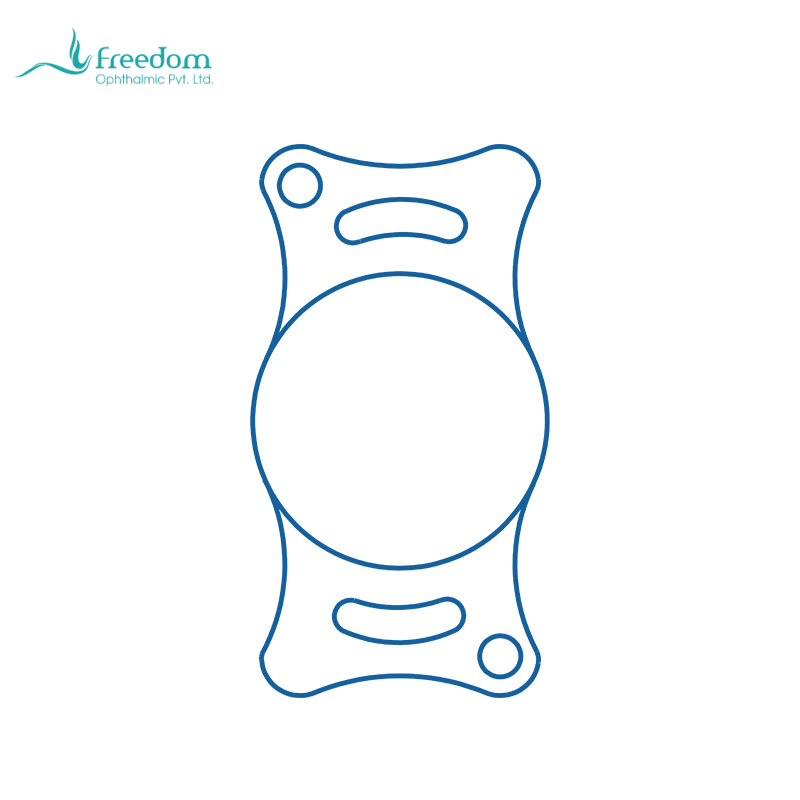
IOL power matters, but it is not the only specification. Whether a veterinary IOL fits a patient also depends on optic diameter, overall length, material, foldability, edge design, and capsular bag conditions. Reducing the decision to canine power or feline power turns an intraocular implant into an ordinary consumable.[1,3]
What does optic diameter affect?
The optic is the central imaging zone. If it is too small, edge-related visual issues may become more likely when the pupil dilates or the IOL is mildly decentered. If it is too large, insertion and capsular fit may become more demanding. Pupil size in dogs and cats is strongly influenced by medication, inflammation, and light, so optic size should be considered together with pupil shape, capsular support, and contraction risk.
Why does overall length matter?
Overall length affects capsular support and centration. A lens that is too short may be unstable; one that is too long may increase capsular tension. Gelatt notes that IOL decentration or luxation can relate to capsular bag size, IOL diameter, excessive capsulotomy size, radial tears, and capsular contraction. Specification choice is therefore part of intraoperative risk management.[1]
How should material and foldability be assessed?
The clinical meaning of hydrophilic acrylic and foldable design lies in implantation behavior. The surgeon should observe loading, controlled entry into the anterior chamber, smooth unfolding, need for manipulation, and capsular traction. A foldable IOL supports the small-incision logic, but if delivery is forced, the minimally invasive advantage declines.
How can a hospital build selection records?
Each case can record model, optic size, overall length, delivery method, repositioning, postoperative centration, pressure, PCO, and owner-observed visual behavior. Over time, the hospital learns which sizes are common, which cases need backup options, and which intraoperative findings tend to change the plan.
Why should canine and feline specifications be read separately?
Dogs and cats differ in globe size, refractive needs, and common case background. IOL specifications should not be interpreted as interchangeable. Canine breed variation often requires a wider range of overall lengths, while feline cases may be more influenced by inflammatory etiology, making ocular environment as important as size.
Purchasing should not ask only about price
When buying IOLs, a veterinary hospital should confirm stable availability of common sizes, packaging and sterilization information, lot traceability, instructions for use, surgeon training support, and emergency replenishment. An intraocular lens is an implant, not an ordinary disposable item.
How can a selection table serve clinical work?
A clinic can build an internal table with species, optic size, overall length, power, material, edge design, practical notes, and inventory. After surgery, the team can add the model used and postoperative findings. The specification table then becomes a clinical learning tool rather than only a purchasing file.
Related product note
The BeneMount brochure lists canine models with 6.00/6.50 mm optic options and 11.00-15.00 mm overall lengths, and feline models with a 7.00 mm optic and 12.00/13.00/14.00 mm overall lengths. The material is hydrophilic acrylic with foldable aspheric design and a 360-degree square edge. These parameters should be matched to patient and capsular conditions.[10]
参考文献
[1] Gelatt KN, Ben-Shlomo G, Gilger BC, Hendrix DVH, Kern TJ, Plummer CE, eds. Veterinary Ophthalmology. 6th ed. Wiley-Blackwell; 2021.
[2] Maggs DJ, Miller PE, Ofri R. Slatter's Fundamentals of Veterinary Ophthalmology. 6th ed. Elsevier; 2018.
[3] British Small Animal Veterinary Association. BSAVA Manual of Canine and Feline Ophthalmology.
[4] Nasisse MP, Davidson MG, Jamieson VE, et al. Phacoemulsification and intraocular lens implantation: a study of technique in 182 dogs. Progress in Veterinary & Comparative Ophthalmology. 1991;1(4):225-238.
[5] Sigle KJ, Nasisse MP. Long-term complications after phacoemulsification for cataract removal in dogs: 172 cases (1995-2002). Journal of the American Veterinary Medical Association. 2006;228(1):74-79.
[6] Maehara S, Itoh N, Wakaiki S, et al. The effects of cataract stage, lens-induced uveitis and cataract removal on ERG in dogs with cataract. Veterinary Ophthalmology. 2007;10(5):308-312.
[7] Bras ID, Colitz CM, Saville WJ, et al. Posterior capsular opacification in diabetic and nondiabetic canine patients following cataract surgery. Veterinary Ophthalmology. 2006;9(5):317-327.
[8] Awasthi N, Guo S, Wagner BJ. Posterior capsular opacification: a problem reduced but not yet eradicated. Archives of Ophthalmology. 2009;127(4):555-562.
[9] Nibourg LM, Gelens E, Kuijer R, Hooymans JMM, van Kooten TG, Koopmans SA. Prevention of posterior capsular opacification. Experimental Eye Research. 2015;136:100-115.
[10] BeneMount. 适应各种需求的犬猫用人工晶体产品彩页. 上海仁山医疗器械有限公司.