

The Cornea Is Not Just a Clear Window
Tonometry estimates intraocular pressure through the corneal interface. Corneal edema, ulceration, dry eye, pigmentation, scarring, mineralization, and irregularity can make measurement harder and interpretation less straightforward.
Rebound tonometry has practical advantages: a small probe, short contact time, and usually no need for topical anesthesia [1]. Slatter also notes that rebound readings may be affected by ocular surface tension, corneal pathology, probe distance, and patient cooperation [2].
Corneal Edema: Ask Why It Is Present
Diffuse corneal haze is a classic sign in glaucoma because high IOP can overwhelm corneal endothelial function. If the eye is painful, the pupil is dilated, episcleral vessels are enlarged, and vision is reduced, a high IOP reading is clinically important.
Corneal edema can also occur with uveitis, endothelial disease, corneal injury, or postoperative change. A normal or low reading does not make the eye routine. Interpret the value with aqueous flare, pupil size, fluorescein staining, endothelial appearance, and the fellow eye.
Corneal Ulceration: Protect the Globe First
With corneal ulcers, the depth and location of the lesion matter. A superficial ulcer in a cooperative patient may still allow careful measurement. A deep ulcer, melting ulcer, impending perforation, or suspicious Seidel test should make any contact procedure very cautious.
If IOP is needed, handling must be gentle and pressure on the eyelids or globe avoided. If the number does not fit the case, repeat it or refer. BSAVA principles for fragile eyes and perioperative handling apply here: the examination should not increase rupture risk [3].
Distance, Angle, and Central Cornea Matter
Rebound tonometry depends on central corneal alignment and appropriate probe distance. Studies show that probe angle, peripheral corneal location, and probe-cornea distance can change readings [4,5]. This matters in corneal disease because the animal may avoid contact and the examiner may drift away from the center to avoid a lesion.
If a nonideal location is used, record it. Do not compare that value as if it were a standard central corneal reading. At recheck, try to use the same device, mode, measurement area, and restraint method.
Turn a Questionable Number into Useful Information
For corneal disease cases, document lesion depth and location, fluorescein result, edema, measurement area, quality indicator, and repeatability. If readings vary widely in the same eye, check technique before changing treatment.
IOP becomes risky when used alone. A reading of 28 mmHg in a painful, edematous eye with a dilated pupil may be urgent. The same number in a scarred cornea with poor alignment and heavy restraint first needs validation.
Related Product Information
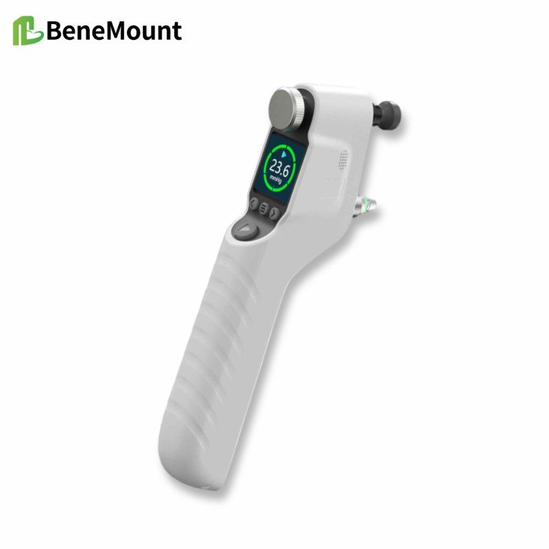
This week’s product materials include the iFalcon V100 Veterinary Tonometer and the Hawkeye V700 veterinary tonometer manuals, quick guides, and brochures. The manuals describe a rebound tonometer for animal IOP measurement using disposable TVP40 probes. During measurement, the probe should be aligned perpendicularly with the central cornea, with the probe tip about 5-7 mm from the corneal surface.
The stated animal modes include feline, canine, equine, lapine, rat, mouse, and primate. The listed measurement range is 5.0-70.0 mmHg, display range 0-99.9 mmHg, and display resolution 0.1 mmHg. For feline, canine, equine, lapine, and primate modes, the stated accuracy is ±1.2 mmHg when IOP is below 20 mmHg and ±2.2 mmHg when IOP is above 20 mmHg; for rats and mice, it is ±10%.
The device forms a final result after six successful measurements, using the best four readings and discarding two outliers according to the manual. Clinically, the number should still be interpreted with the case context: signs, corneal status, anterior chamber findings, medications, restraint, species mode, and repeatability.
References
Gelatt KN, Gilger BC, Kern TJ, et al. Veterinary Ophthalmology. 6th ed. Wiley-Blackwell; 2021. Chapters on ophthalmic examination, tonometry, canine glaucoma, and anterior uveal disease.
Maggs DJ, Miller PE, Ofri R. Slatter's Fundamentals of Veterinary Ophthalmology. 4th ed. Saunders Elsevier; 2008. Chapters on ophthalmic examination, tonometry, uveitis, and glaucoma.
Gould D, McLellan G, eds. BSAVA Manual of Canine and Feline Ophthalmology. 3rd ed. BSAVA; 2014. Chapters on ocular examination, uveal tract disease, glaucoma, and problem-oriented presentations.
de Oliveira JK, Montiani-Ferreira F, Williams DL. The influence of the tonometer position on canine intraocular pressure measurements using the Tonovet rebound tonometer. Open Vet J. 2018;8(1):68-76. PMID: 29721435.
Rodrigues BD, Montiani-Ferreira F, Bortolini M, Somma AT, Komáromy AM, Dornbusch PT. Intraocular pressure measurements using the TONOVET rebound tonometer: influence of the probe-cornea distance. Vet Ophthalmol. 2021;24 Suppl 1:175-185. PMID: 33070466. DOI: 10.1111/vop.12832.