

Do Not Record Only One Number
A note that says “left eye 32” is not enough. It does not state the unit, device, species mode, repeatability, corneal status, restraint, fellow-eye value, or how the number fits the signs.
Gelatt notes that outpatient tonometry is a snapshot, but repeat tonometry is invaluable for monitoring disease and response to therapy [1]. The purpose of recording IOP is not to store a number; it is to preserve comparable clinical evidence.
What a Useful IOP Entry Includes
A useful entry includes OD/OS, value in mmHg, device type, species mode, quality indicator, number of repeats, disposable probe use, medications given before measurement, fluorescein result, corneal edema or scarring, restraint, and patient position.
For glaucoma, uveitis, postoperative, and corneal disease cases, also record pupil status, visual function, pain, aqueous flare, corneal edema, current medication, and recheck timing. At the next visit, this helps separate disease change from measurement-condition change.
Keep Follow-Up Comparable
Readings from different devices and modes should not be treated as interchangeable. A 2024 multi-species study found significant differences between rebound tonometer models in several species, supporting device-specific reference values and follow-up interpretation [4].
If the device changes between clinics, say so in the record. For follow-up, try to keep the same device, species mode, time of day, restraint method, and measurement area. In chronic glaucoma and postoperative eyes, inconsistent conditions can obscure the trend.
Make Referral Notes Clinically Useful
A strong referral note includes onset, signs, eye, serial IOP values, highest and lowest values, fellow-eye status, fluorescein result, anterior chamber findings, medications and response, suspected lens disease or cataract, and whether vision has declined.
Do not write only “suspected glaucoma.” For low-IOP red eye, write “suspected anterior uveitis; needs cause assessment and secondary glaucoma monitoring.” For ulceration with high IOP, include measurement conditions and ulcer depth so the ophthalmologist can judge reliability and priority.
Turn Quality Control into a Habit
Clinics can build an IOP recheck template. A nurse or assistant prepares the device, confirms disposable probes and species mode, and the veterinarian interprets the value and plan. The medical record requires eye, unit, device, and repeat result.
This reduces missed measurements and non-comparable records. For a veterinary practice, the value of a tonometer is not only the device; it is the more consistent pathway it creates for red eye, glaucoma, uveitis, and postoperative care.
Related Product Information
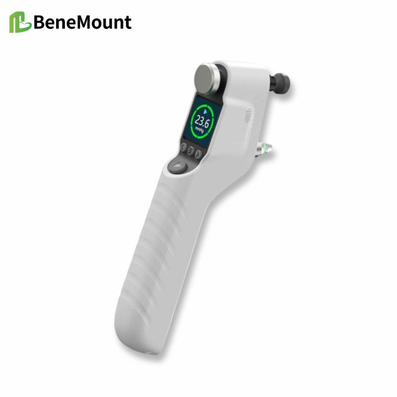
This week’s product materials include the iFalcon V100 Veterinary Tonometer and the Hawkeye V700 veterinary tonometer manuals, quick guides, and brochures. The manuals describe a rebound tonometer for animal IOP measurement using disposable TVP40 probes. During measurement, the probe should be aligned perpendicularly with the central cornea, with the probe tip about 5-7 mm from the corneal surface.
The stated animal modes include feline, canine, equine, lapine, rat, mouse, and primate. The listed measurement range is 5.0-70.0 mmHg, display range 0-99.9 mmHg, and display resolution 0.1 mmHg. For feline, canine, equine, lapine, and primate modes, the stated accuracy is ±1.2 mmHg when IOP is below 20 mmHg and ±2.2 mmHg when IOP is above 20 mmHg; for rats and mice, it is ±10%.
The device forms a final result after six successful measurements, using the best four readings and discarding two outliers according to the manual. Clinically, the number should still be interpreted with the case context: signs, corneal status, anterior chamber findings, medications, restraint, species mode, and repeatability.
References
Gelatt KN, Gilger BC, Kern TJ, et al. Veterinary Ophthalmology. 6th ed. Wiley-Blackwell; 2021. Chapters on ophthalmic examination, tonometry, canine glaucoma, and anterior uveal disease.
Maggs DJ, Miller PE, Ofri R. Slatter's Fundamentals of Veterinary Ophthalmology. 4th ed. Saunders Elsevier; 2008. Chapters on ophthalmic examination, tonometry, uveitis, and glaucoma.
Gould D, McLellan G, eds. BSAVA Manual of Canine and Feline Ophthalmology. 3rd ed. BSAVA; 2014. Chapters on ocular examination, uveal tract disease, glaucoma, and problem-oriented presentations.
Kovalcuka L, Mālniece A, Vanaga J. Comparison of Tonovet and Tonovet Plus tonometers for measuring intraocular pressure in dogs, cats, horses, cattle, and sheep. Vet World. 2024;17(2):384-388. PMID: 38595645.