

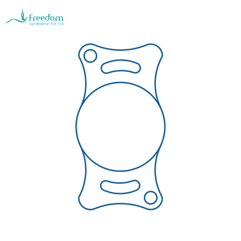
A veterinary intraocular lens is an optical implant placed during cataract surgery, usually in the capsular bag, to replace part of the refractive function of the removed crystalline lens. Cataract surgery in dogs and cats is not simply the removal of an opacity. If the lens is removed without an implant, the eye may become clear but aphakic, leaving a major refractive difference from a normal eye.[1,2]
Why consider an IOL after cataract removal?
An aphakic animal may perceive light and large objects, yet still struggle with near judgment, stairs, jumping, moving targets, and low-light activity. Dogs and cats do not describe blur or glare. Clinicians judge function through behavior: confidence on stairs, accuracy when jumping onto furniture, obstacle avoidance, and toy tracking. In suitable cases, the IOL helps focus the restored optical pathway onto the retina, converting clarity into usable vision.[1-3]
Which cases deserve an IOL discussion?
Good candidates usually share several features: controlled intraocular inflammation, stable pressure, corneal and tear-film health compatible with recovery, retinal potential, acceptable capsular support, and an owner able to medicate and return for rechecks. If posterior capsule rupture, zonular instability, active uveitis, or poor retinal prognosis is present, ocular safety should take priority over primary implantation.[1,2]
How should the surgeon explain it?
The IOL is best described as an implant that helps restore focus, not as an optional luxury upgrade. Cataract removal clears the obstruction; the IOL reduces the refractive deficit of aphakia. Owners should also be told before surgery that final implantation depends on capsular and intraocular conditions. If the risk is too high, leaving the eye without a primary IOL may be the safer decision.
What should be checked after surgery?
Follow-up should not stop at corneal clarity. The record should include anterior chamber inflammation, intraocular pressure, IOL centration, pupil shape, posterior capsule clarity, and owner-observed visual behavior. This separates a cosmetically clear eye from a functionally successful eye. Long-term rechecks are especially important for PCO, pressure elevation, and IOL displacement.[5,7]
Common mistake: cataract removal equals vision recovery
Cataract removal solves the problem of light being blocked by an opaque lens. It does not guarantee useful vision. Postoperative function also depends on retinal health, pressure, inflammation, IOL position, and posterior capsule clarity. If the eye looks clear but the animal still hesitates or bumps into objects, the clinician should reassess posterior segment status and optical function rather than simply saying recovery is slow.
How can functional vision be assessed before surgery?
Ask specific questions: Does the animal go down stairs confidently? Can it jump onto a familiar surface accurately? Does it hesitate in dim light? Can it track toys or food? Does it walk along walls? Recording these behaviors before surgery and asking the same questions after surgery is more useful than asking whether the pet can see.
How can clinics build an IOL case record?
Useful fields include species, breed, age, cataract stage, diabetic status, preoperative pressure, LIU status, IOL implantation, IOL model, postoperative pressure peak, posterior capsule status, and owner feedback. Over time, these records help the clinic identify which cases need early referral, which lens options are used most often, and which complications deserve more attention.
Related product note
BeneMount veterinary intraocular lenses are described as hydrophilic acrylic, aspheric, and foldable. The brochure lists +41.00D canine and +53.50D feline models with species-specific optic and overall-length options. These specifications support inventory and intraoperative selection, but they do not replace case selection or surgeon judgment.[10]