

Owners often describe cataracts as a white eye, but for veterinarians cataract is not merely an appearance. It is a clinical condition involving lens opacity, intraocular inflammation, retinal function, and often systemic disease. The therapeutic value of cataract surgery depends on whether the animal still has recoverable visual potential and whether intervention occurs before inflammation and secondary complications narrow the window for success. Veterinary ophthalmology references classify cataracts as incipient, immature, mature, and hypermature; these stages are clinically meaningful because they influence surgical difficulty, lens-induced uveitis, and prognosis.[1-3]
An incipient cataract may affect only a small portion of the lens, whereas a mature cataract can cause profound visual impairment. A hypermature or Morganian cataract is not simply a whiter lens. It reflects liquefaction of lens proteins, capsular permeability changes, and increased antigenic stimulation within the eye. Lens-induced uveitis, posterior synechiae, secondary glaucoma, and retinal complications become more important. If chronic inflammation or retinal dysfunction is already present, removing the cataract alone may not restore meaningful vision.[1,2]
Diabetic dogs deserve particular attention. Diabetic cataracts can progress rapidly because osmotic changes within the lens promote water influx and swelling. Owners may present only after the dog collides with furniture, avoids stairs, or becomes reluctant in dim light, but by that stage inflammation may already be limiting the surgical window. For diabetic patients, cataract surveillance should be part of diabetes follow-up rather than a discussion deferred until blindness is complete.[1,2,4]
Preoperative assessment should answer four questions. Is the opacity sufficient to explain visual loss? Are inflammation and intraocular pressure controlled? Is the retina likely to function after media clarity is restored? Can the owner complete frequent topical medication and rechecks? Slit-lamp examination, tonometry, corneal and tear-film assessment are foundational. When the fundus cannot be visualized, ocular ultrasonography and electroretinography can be important for selection. In cats, clinicians should also look for uveitis, trauma, FIP-associated ocular disease, or other systemic causes because feline cataract etiologies differ from canine patterns.[1,2]
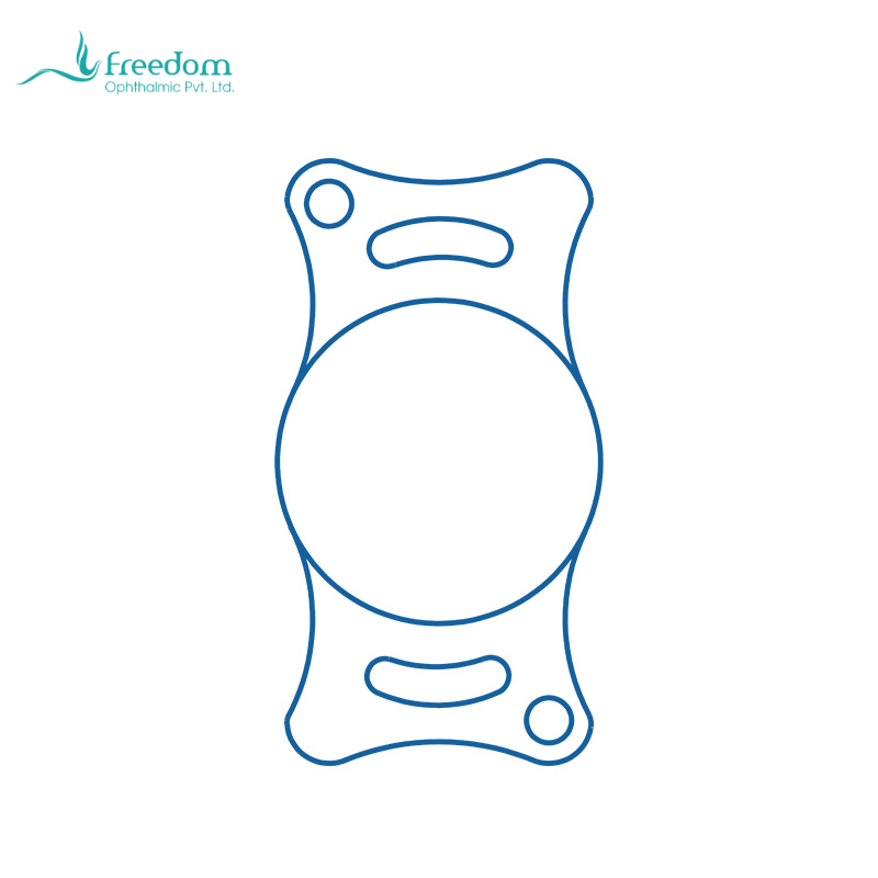
The objective of cataract surgery is not a clear-looking eye; it is useful vision with long-term ocular stability. Phacoemulsification allows small-incision lens removal while preserving the capsular bag, which provides the anatomic basis for intraocular lens placement. A veterinary intraocular lens reduces the major refractive deficit of aphakia and can improve functional vision for daily activity. The decision to implant a canine or feline IOL should integrate preoperative findings, capsular support, inflammation status, and owner compliance.[1,2,5]
Cataract stage also changes how the case should be discussed with owners. When a cataract is incipient or immature, owners may believe the animal still sees well enough and delay evaluation. When it is mature or hypermature, they may assume it is too late. The veterinarian should shift the conversation from whiteness to risk: Is inflammation present? Has intraocular pressure changed? Can the retina be evaluated? Will waiting increase capsular or iris complications? Timing should be based on ocular environment and visual potential, not simply on how white the lens appears.[1-3]
In cats, cataracts should rarely be separated from etiology. Primary age-related cataract is less typical than in many human patients; uveitis, trauma, lens instability, nutritional problems, and systemic disease may all be relevant. If active uveitis is missed, IOL implantation may face a higher inflammatory burden. The preoperative question in feline cataract is therefore not only whether the lens can be removed, but why the cataract developed.[1,2]
For referral systems, primary practices can classify cataract cases into three groups: early cases that can be monitored with scheduled rechecks, progressive cases that should be assessed by an ophthalmologist soon, and high-risk cases with pain, inflammation, abnormal pressure, or an unviewable fundus. This classification reduces delay and gives the referral center a clearer disease timeline.
A common scenario is the diabetic dog whose lens changes rapidly from mild opacity to bilateral mature cataracts. Owners may initially interpret the change as aging. If the primary clinician only recommends observation until the eyes become red, pressure fluctuates, or synechiae develop, surgical difficulty has already increased. A better approach is to establish ophthalmic surveillance when diabetes is diagnosed and refer promptly when lens change accelerates. This is not excessive care; it protects retinal vision that may still be recoverable.
Another scenario is unilateral cataract in a cat. The owner may notice only a color change in the pupil, but the veterinarian should look for uveitis or trauma. If the cause is not investigated and the case is treated simply as a surgical cataract, postoperative inflammatory risk may be underestimated. In feline cataract cases, etiologic investigation and inflammation control often come before implant selection.