

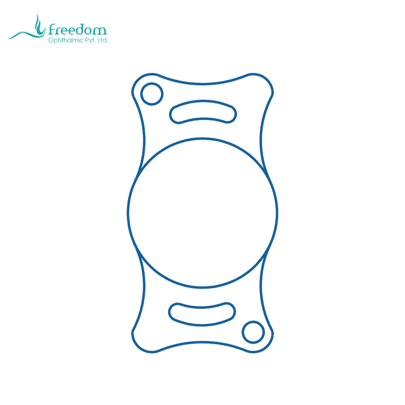
After cataract extraction, the ocular media may become clear, but the optical system is not automatically restored. The crystalline lens contributes substantially to the refractive power of the eye. Without an intraocular lens, the patient is aphakic and typically left with a major hyperopic refractive deficit. The animal may perceive light and large objects but still struggle with spatial judgment, stairs, jumping, moving targets, and close interaction. This optical problem is the fundamental reason to discuss IOL implantation.[1,2]
Human cataract surgery historically used thick spectacles or contact lenses to correct aphakia; modern cataract surgery usually uses an intraocular lens. Veterinary patients cannot reliably wear high-power spectacles and are poor long-term candidates for aphakic contact lens correction. Implanting a canine or feline IOL during surgery is therefore a logical route to functional visual rehabilitation. In animals, visual quality is observed through behavior: confidence on stairs, accuracy of jumping, obstacle avoidance, tracking of toys, and activity in dim light.[1,2]
An IOL also reduces the burden of postoperative adaptation. Animals cannot understand that the eye looks quiet but vision remains optically defocused. Owners may interpret hesitation or bumping into objects as surgical failure. In suitable eyes, in-the-bag IOL implantation brings the refractive state closer to physiologic function, making behavioral improvement easier to observe. The goal is not to promise normal vision; it is to reduce the optical handicap of aphakia and convert surgical clarity into usable vision.[1,2]
IOL power cannot be borrowed directly from human assumptions. Dogs and cats differ in axial length, corneal curvature, anterior chamber depth, and native lens power. Standard veterinary IOLs can meet functional targets in many routine cases, while special cases benefit from biometry and refractive planning. Effective lens position, capsular contraction, haptic support, and postoperative inflammation also influence the final refractive result, so labeled diopter is only one part of the outcome.[1,2,9]
Not every cataract patient should receive an IOL. Posterior capsule rupture, zonular instability, severe uveitis, uncontrolled intraocular pressure, or poor retinal prognosis can shift priorities. The clinician must balance optical benefit against intraocular safety. A mature surgical plan includes an ideal plan for in-the-bag implantation, a contingency plan for altered support or lens choice, and a safety plan in which primary implantation is abandoned if ocular stability requires it.
Optically, the value of an IOL is not merely that it has a labeled power. After lens removal, the eye loses a major refractive element located behind the iris, and light no longer focuses in the same way. The IOL must remain relatively stable in the capsular bag for postoperative refraction to be predictable. If the lens decentrates, tilts, or is affected by capsular contraction, visual quality may suffer even when the cornea is clear and pressure is normal. Postoperative evaluation of IOL position and capsular status is therefore as important as assessment of the anterior chamber.[1,2]
Visual assessment in dogs and cats requires behavioral context. Animals do not report distance clarity, near blur, or glare, but owners can observe route choice, confidence when jumping, response to moving objects, and activity in dim light. Veterinarians can record these functional behaviors before surgery and ask the same questions at rechecks. Serial functional observation is more useful than a single owner impression.
If an IOL is not implanted, the expectation should still be explained. Aphakia does not necessarily mean surgical failure; in some eyes, leaving the patient aphakic is safer than placing an implant in a compromised eye. Owners should understand, however, that the animal may retain a refractive deficit. Explaining why an eye is not implanted is as important as explaining why another eye is a good candidate.
A simple analogy can help owners understand the optics. Cataract removal clears fog from the camera, but without an appropriate focusing lens the image may still be blurred. The IOL helps focus the restored optical pathway onto the retina. This explanation helps owners understand why IOL implantation is discussed during surgery and helps referring veterinarians present the implant as part of visual rehabilitation rather than as decoration.
Communication is equally important when primary implantation should be abandoned. If the posterior capsule ruptures, continuing to pursue an implant may increase vitreous disturbance, inflammation, and later complications. In that situation, preserving intraocular stability is more important than achieving ideal refraction. If this possibility is included in informed consent before surgery, owners are more likely to understand an intraoperative change of plan.