

Red and Blue Pupillary Responses: Why Color Can Add Clinical Clues
Why not use only a white penlight?
A white penlight is adequate for basic PLR testing, but it produces a mixed response across multiple wavelengths. Red and blue stimuli can make the observation more structured.
Outer retinal rods and cones contribute to image-forming vision and influence the pupil. Intrinsically photosensitive retinal ganglion cells containing melanopsin are especially sensitive to short-wavelength blue light and contribute to sustained pupil constriction and non-image-forming visual responses [6,7].
This does not mean that blue light diagnoses one disease and red light diagnoses another. The practical value is that different stimuli help the veterinarian observe onset, amplitude, sustained constriction, pupil escape, and asymmetry more consistently.
What can a red-light response suggest?
Red-light testing is often discussed in relation to outer retinal photoreceptor function. When a dog has progressive night blindness, dilated pupils, increased tapetal reflection, narrowed retinal vessels, or breed risk for inherited retinopathy, a weak red-light response increases concern for retinal disease.
In progressive retinal atrophy and related inherited retinal disorders, early clinical signs, fundus changes, and pupil responses do not always appear at the same time. Veterinary texts emphasize that confirmation may require fundus examination, genetic testing, electroretinography, or follow-up [1,2].
The red-light response is therefore a screening clue. It warns the clinician not to reduce the case to a corneal or lens problem, but it does not replace ERG or identify every retinal disorder by itself.
Why does blue light attract attention?
Blue light is closely linked to melanopsin-mediated pathways. In mammals, melanopsin retinal ganglion cells contribute to sustained pupil constriction and circadian light responses. In severe outer retinal disease, some blue-light driven slow responses may remain, which is why red-blue comparison can be clinically informative [6,7].
In canine sudden acquired retinal degeneration syndrome, dogs may become blind abruptly, the fundus may look normal early, and ERG is commonly used to confirm severe outer retinal dysfunction. A residual light reflex does not rule out serious retinal disease [8].
A poor blue-light response can also occur with optic nerve or more proximal pathway disease. The pupillary signal still has to travel through the optic nerve and midbrain, so color testing never removes the need for neuro-ophthalmic localization.
What is pupil escape?
Pupil escape describes initial constriction followed by gradual redilation while illumination continues. It can suggest poor maintenance of the reflex and may raise concern for retinal, optic nerve, or central pathway dysfunction.
The product chart supplied this week highlights the pattern of initial constriction followed by dilation during continued illumination. Clinically, this dynamic behavior should be recorded directly instead of being reduced to “PLR present.”
Technique matters. Light movement, head movement, changing room light, excessive exposure time, and patient stress can all mimic abnormal dynamics. A finding becomes more useful when it repeats under similar conditions and agrees with the rest of the examination.
Common mistakes when using red and blue light
Do not treat a color-response chart as a diagnosis table. Diseases overlap, and one disease can look different at different stages.
Do not ignore ocular media opacity. Corneal edema, aqueous flare, cataract, and vitreous opacity can change the light reaching the retina.
Do not isolate the test from the rest of the workflow. Red-blue PLR is best interpreted alongside history, visual behavior, menace response, dazzle reflex, slit lamp examination, tonometry, fundus examination, and ERG when indicated.
Related Product Information
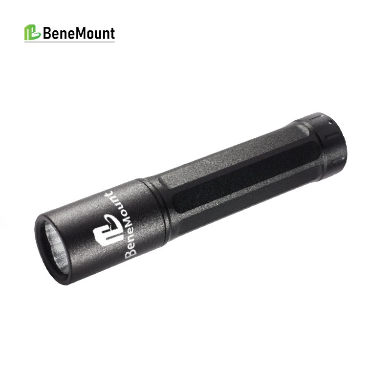
The CPRL Tester uses red and blue light stimulation and is accompanied by a disease-response comparison chart. For veterinary hospitals, its practical value is standardization: a more consistent light source and observation language for screening, follow-up, and referral communication.
References / 参考文献
[1] Gelatt KN, Gilger BC, Kern TJ, eds. Veterinary Ophthalmology. 5th ed. Wiley-Blackwell; 2013. Local reference PDF: Kirk N. Gelatt – Veterinary Ophthalmology Two-Volume Set.
[2] Maggs DJ, Miller PE, Ofri R. Slatter's Fundamentals of Veterinary Ophthalmology. 4th ed. Saunders Elsevier; 2008. Local reference PDF.
[3] Gould D, McLellan G, eds. BSAVA Manual of Canine and Feline Ophthalmology. BSAVA; local reference PDF.