

Blue-Light Pupillary Response and Melanopsin: Why It Is Not the Same as Vision
Why blue light is discussed separately
Many clinics still use only a white penlight. Blue-light testing became more clinically interesting as intrinsically photosensitive retinal ganglion cells were characterized. These cells contain melanopsin, are sensitive to short-wavelength light, and contribute to sustained pupil constriction, circadian entrainment, and other non-image-forming light responses [6,7,8].
The practical lesson is important: the pupil reflex is not identical to image-forming vision. Even when outer retinal photoreceptors are severely damaged, some inner retinal light responses may remain.
Why a blue response does not prove vision
Vision requires image formation through the cornea, lens, vitreous, photoreceptors, bipolar cells, ganglion cells, optic nerve, visual pathways, and cortex. A sustained blue-light pupil response mainly shows activity within non-image-forming light pathways and the reflex arc. It does not prove that the animal can identify objects, judge distance, or navigate safely.
Clinicians may see animals with poor menace response or poor obstacle navigation that still have a residual PLR. Cortical blindness can also spare PLR. Veterinary texts are consistent on this point: PLR is a localization sign, not a direct measure of vision [1,2].
Cases that are easy to misread
SARDS is a key example. Dogs may become suddenly blind, have an unremarkable early fundus, and show severe outer retinal dysfunction on ERG, yet the pupil reflex may not be completely absent. If a residual PLR is interpreted as “retinal function is acceptable,” the workup can go in the wrong direction [9].
Advanced PRA, optic nerve disease, severe uveitis, glaucoma, and central lesions can also produce mismatches between pupil response and visual behavior. Red and blue testing describes a pattern; it does not replace fundus examination, IOP, ERG, or neuroimaging.
How to document blue-light findings
Avoid writing only “blue positive.” Describe latency, amplitude, duration, asymmetry, and pupil escape. For example: “blue stimulus produced mild constriction after one second; constriction was maintained for five seconds; right eye weaker than left.”
When red light is also used, state whether the red response is weaker or shorter. This helps the next clinician judge whether outer retinal and inner retinal light responses appear different.
Practical boundaries for clinics
Blue-light testing is useful for screening, follow-up, and referral documentation, especially when behavior testing is unreliable, the fundus is atypical, or serial comparison matters.
The boundary should be explicit: a blue response is not proof of useful vision, not a rule-out test for SARDS, and not a replacement for ERG. Its value is standardized observation and better clinical questioning.
Related Product Information
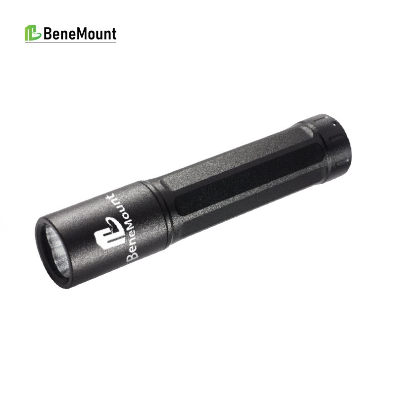
The CPRL Tester’s red and blue light materials can help clinics record blue-light responses more consistently. Results should be integrated into a complete ophthalmic record rather than treated as a stand-alone diagnosis.
References
[1] Gelatt KN, Gilger BC, Kern TJ, eds. Veterinary Ophthalmology. Wiley-Blackwell. Local reference PDF.
[2] Maggs DJ, Miller PE, Ofri R. Slatter's Fundamentals of Veterinary Ophthalmology. Saunders/Elsevier. Local reference PDF.
[3] Gould D, McLellan GJ, eds. BSAVA Manual of Canine and Feline Ophthalmology. BSAVA. Local reference PDF.
[4] Companion Animal Ophthalmology. Local Chinese reference PDF.
[5] Small Animal Ophthalmology. Local Chinese reference PDF.
[6] Hattar S, Liao HW, Takao M, Berson DM, Yau KW. Melanopsin-containing retinal ganglion cells: architecture, projections, and intrinsic photosensitivity. Science. 2002;295(5557):1065-1070.
[7] Berson DM, Dunn FA, Takao M. Phototransduction by retinal ganglion cells that set the circadian clock. Science. 2002;295(5557):1070-1073.
[8] Do MTH, Yau KW. Intrinsically photosensitive retinal ganglion cells. Physiological Reviews. 2010;90(4):1547-1581.
[9] Komaromy AM, Abrams KL, Heckenlively JR, et al. Sudden acquired retinal degeneration syndrome (SARDS): a review and proposed strategies toward a better understanding of pathogenesis, early diagnosis, and therapy. Veterinary Ophthalmology. 2016;19(4):319-331.
[10] Hall CA, Chilcott RP. Eyeing up the future of the pupillary light reflex in neurodiagnostics. Diagnostics. 2021;11(10):1795.
[11] Product files for this week: CPRL Tester, red/blue light manual PDF, and pupil reaction chart image. Product files were used only to verify product name and visible use cues.