

Baseline IOP Reduces Guesswork
Before ophthalmic procedures, baseline IOP is often more useful than expected. Cataract, lens instability, anterior uveitis, glaucoma-risk breeds, trauma, and deep corneal disease may all show pressure changes during anesthesia, dilation, surgery, or postoperative inflammation.
Gelatt and BSAVA emphasize IOP assessment as part of ophthalmic examination and follow-up [1,3]. Without a preoperative value, a postoperative reading of 26 mmHg or 8 mmHg is harder to interpret. It may be new, pre-existing, or partly related to handling and drugs.
Anesthesia and Handling Change the Number
Many sedatives and general anesthetics reduce IOP, while ketamine may transiently increase it. Stress, struggling, neck pressure, lid traction, and pressure on the globe may also alter the reading [2,3]. Peri-anesthetic tonometry must therefore be interpreted with restraint and drug context.
BSAVA cautions that fragile eyes should be protected from perioperative increases in IOP, especially deep ulcers and eyes at risk of rupture [3]. In practice, allow the patient to settle, avoid tight collars, and use controlled assistance or a harness when appropriate.
Postoperative Ocular Hypertension Must Be Found Early
After cataract surgery, glaucoma procedures, intraocular inflammation, and lens-related disease, increased IOP can affect comfort and visual prognosis. Slatter describes postoperative IOP spikes after some glaucoma procedures that may persist for days and require close follow-up [2].
A postoperative check should not stop at corneal clarity and wound appearance. Record IOP, anterior chamber inflammation, pupil status, corneal edema, fundus visibility, and medication compliance. If IOP rises with pain or reduced vision, contact a veterinary ophthalmologist promptly.
Low IOP Can Also Signal Trouble
Postoperative hypotony may reflect wound leakage, ciliary body inflammation, uncontrolled intraocular inflammation, or structural compromise. Mild transient reduction may fit the procedure; marked hypotony, shallow anterior chamber, suspected leakage, or a soft globe requires urgent attention.
In uveitis, a low IOP that gradually returns toward the fellow eye may indicate improvement. If it then climbs above normal or above the fellow eye, secondary glaucoma becomes a concern. The trend is more informative than one isolated measurement.
Build Practical Recheck Points
Clinics can place IOP directly into the perioperative checklist: preoperative baseline, pre-anesthetic or post-sedation value, early postoperative value, 24-48 hour recheck, one-week recheck, and more frequent checks for high-risk eyes. Each entry should include device, eye, species mode, quality indicator, and medication status.
The goal is not paperwork. It is to help the team decide when to refer, add medication, taper medication, or re-examine the cornea and anterior chamber. Ophthalmic surgical quality control depends on comparable data over time.
Related Product Information
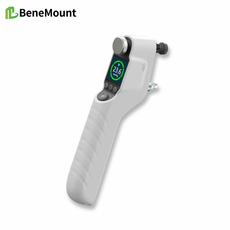
This week’s product materials include the iFalcon V100 Veterinary Tonometer and the Hawkeye V700 veterinary tonometer manuals, quick guides, and brochures. The manuals describe a rebound tonometer for animal IOP measurement using disposable TVP40 probes. During measurement, the probe should be aligned perpendicularly with the central cornea, with the probe tip about 5-7 mm from the corneal surface.
The stated animal modes include feline, canine, equine, lapine, rat, mouse, and primate. The listed measurement range is 5.0-70.0 mmHg, display range 0-99.9 mmHg, and display resolution 0.1 mmHg. For feline, canine, equine, lapine, and primate modes, the stated accuracy is ±1.2 mmHg when IOP is below 20 mmHg and ±2.2 mmHg when IOP is above 20 mmHg; for rats and mice, it is ±10%.
The device forms a final result after six successful measurements, using the best four readings and discarding two outliers according to the manual. Clinically, the number should still be interpreted with the case context: signs, corneal status, anterior chamber findings, medications, restraint, species mode, and repeatability.
References
Gelatt KN, Gilger BC, Kern TJ, et al. Veterinary Ophthalmology. 6th ed. Wiley-Blackwell; 2021. Chapters on ophthalmic examination, tonometry, canine glaucoma, and anterior uveal disease.
Maggs DJ, Miller PE, Ofri R. Slatter's Fundamentals of Veterinary Ophthalmology. 4th ed. Saunders Elsevier; 2008. Chapters on ophthalmic examination, tonometry, uveitis, and glaucoma.
Gould D, McLellan G, eds. BSAVA Manual of Canine and Feline Ophthalmology. 3rd ed. BSAVA; 2014. Chapters on ocular examination, uveal tract disease, glaucoma, and problem-oriented presentations.
Gelatt KN, Gilger BC, Kern TJ, et al. Veterinary Ophthalmology. 6th ed. Wiley-Blackwell; 2021. Chapters on pharmacology, anesthesia, diagnostics, and canine glaucoma.