

Do Not Label Every Red Eye as Conjunctivitis
The most common mistake in canine and feline red eye is to treat every red, tearing, squinting eye as conjunctivitis. Conjunctivitis is common, but glaucoma, anterior uveitis, corneal ulceration, scleritis, orbital disease, and lens luxation can all begin with a similar owner complaint.
Tonometry helps separate these paths. Slatter and Gelatt both describe IOP measurement as more than a glaucoma test; it is part of the workup for red, painful, cloudy, and inflamed eyes [1,2]. A dog with acute blepharospasm, corneal edema, a dilated pupil, and high IOP is a very different patient from a cat with miosis, aqueous flare, and low IOP.
How IOP Changes the First-Visit Plan
A practical first-visit sequence is history, visual behavior, pupils, corneal clarity, conjunctiva, anterior chamber assessment, then IOP measurement without pressure on the neck, eyelids, or globe. Fluorescein staining, slit-lamp examination, and posterior segment assessment can then be added according to the findings.
If IOP is clearly increased, especially with pain, corneal edema, mydriasis, or vision loss, the case should be treated as urgent glaucoma or referred quickly. Gelatt notes that normal IOP in most animals is roughly in the 15-25 mmHg range, although species, age, device, and study design matter. A difference of more than about 8 mmHg between fellow eyes should trigger a careful search for disease [1].
Low IOP Is Also a Diagnostic Finding
Low IOP in a red eye is easy to miss. Anterior uveitis disrupts the blood-aqueous barrier and often reduces aqueous humor production. Clinically, the eye may show ciliary injection, miosis, aqueous flare, keratic precipitates, photophobia, and hypotony [2,3].
A low reading does not make the case mild. Uveitis can lead to secondary glaucoma, synechiae, cataract progression, and posterior segment disease. A baseline IOP at the first visit helps the clinician judge whether inflammation is improving or whether secondary ocular hypertension is developing.
Protect the Patient and the Reading
Red-eye patients are often painful and anxious. Rough restraint can change the number. BSAVA specifically cautions against pressure on the neck, eyelids, or globe during tonometry because it may falsely increase IOP [3]. Deep ulcers, fragile globes, and recent intraocular surgery require especially careful handling.
When a number does not fit the eye, repeat the measurement. Check central corneal alignment, distance, species mode, and quality indicator. Tonometry is a key piece of evidence, not the whole diagnosis.
A Practical Habit for General Practice
Making IOP a routine part of red-eye triage improves decision-making. Normal IOP does not rule out corneal ulceration, conjunctivitis, or early uveitis. High IOP does not prove primary glaucoma by itself. Low IOP should not be ignored.
The medical record should include OD/OS, the value, device type, species mode, restraint, corneal status, staining result, and any topical medication already given. At recheck, the trend often matters more than a single number.
Related Product Information
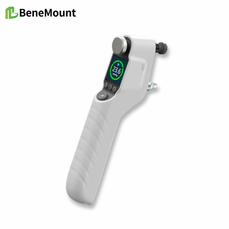
This week’s product materials include the iFalcon V100 Veterinary Tonometer and the Hawkeye V700 veterinary tonometer manuals, quick guides, and brochures. The manuals describe a rebound tonometer for animal IOP measurement using disposable TVP40 probes. During measurement, the probe should be aligned perpendicularly with the central cornea, with the probe tip about 5-7 mm from the corneal surface.
The stated animal modes include feline, canine, equine, lapine, rat, mouse, and primate. The listed measurement range is 5.0-70.0 mmHg, display range 0-99.9 mmHg, and display resolution 0.1 mmHg. For feline, canine, equine, lapine, and primate modes, the stated accuracy is ±1.2 mmHg when IOP is below 20 mmHg and ±2.2 mmHg when IOP is above 20 mmHg; for rats and mice, it is ±10%.
The device forms a final result after six successful measurements, using the best four readings and discarding two outliers according to the manual. Clinically, the number should still be interpreted with the case context: signs, corneal status, anterior chamber findings, medications, restraint, species mode, and repeatability.
References
Gelatt KN, Gilger BC, Kern TJ, et al. Veterinary Ophthalmology. 6th ed. Wiley-Blackwell; 2021. Chapters on ophthalmic examination, tonometry, canine glaucoma, and anterior uveal disease.
Maggs DJ, Miller PE, Ofri R. Slatter's Fundamentals of Veterinary Ophthalmology. 4th ed. Saunders Elsevier; 2008. Chapters on ophthalmic examination, tonometry, uveitis, and glaucoma.
Gould D, McLellan G, eds. BSAVA Manual of Canine and Feline Ophthalmology. 3rd ed. BSAVA; 2014. Chapters on ocular examination, uveal tract disease, glaucoma, and problem-oriented presentations.
von Spiessen L, Karck J, Rohn K, Meyer-Lindenberg A. Clinical evaluation of the Tonovet rebound tonometer in dogs and cats considering potential errors in handling. Tierarztl Prax Ausg K Kleintiere Heimtiere. 2013;41(4):213-220. PMID: 23958704.