

Why IOP Testing in Dogs and Cats Should Not Wait Until Glaucoma Is Obvious
What question does IOP answer?
Intraocular pressure is more than a number in mmHg. It reflects the balance between aqueous humor production and outflow, and it is influenced by ciliary body function, the iridocorneal angle, lens position, and intraocular inflammation. Standard veterinary ophthalmology texts place tonometry within the core eye examination, especially for glaucoma, uveitis, lens luxation, trauma, and postoperative monitoring [1,2,3].
Dogs and cats rarely show ocular pain in a neat way. The owner may report squinting, tearing, redness, blue cornea, reluctance to be touched around the head, or simply reduced activity. Without tonometry, high IOP may be missed, and low IOP from uveitis may be mistaken for uncomplicated conjunctivitis.
IOP therefore connects history, pain, corneal clarity, anterior chamber findings, pupil size, lens position, and fundus assessment. The number does not diagnose alone, but it changes the urgency and direction of the next step.
Which cases deserve active IOP measurement?
Red and painful eyes should be measured unless the eye is too fragile to touch. Glaucoma, anterior uveitis, corneal ulceration with intraocular reaction, and trauma can all alter IOP. High pressure raises concern for optic nerve and retinal risk, while low pressure often points toward inflammation or reduced ciliary body function.
Corneal edema, mydriasis, buphthalmos, and sudden visual change are also strong reasons to measure. If these signs accompany high IOP, the case should move quickly into a glaucoma pathway rather than waiting for a recheck days later [1,2].
IOP is also important for rechecks and preoperative workups. Cataract patients, lens luxation cases, uveitis, glaucoma treatment, and postoperative eyes all benefit from serial records. Trends are often more useful than one isolated number.
How to interpret one pressure reading
Interpret IOP by species, side-to-side difference, and clinical context. Normal ranges vary with species, device, time of day, age, technique, and study design. In practice, comparing both eyes and matching the number to the ocular findings is safer than treating one threshold as absolute.
A mildly high reading does not automatically mean glaucoma. Stress, neck pressure during restraint, eyelid squeezing, poor probe alignment, corneal disease, and poor repeatability can all distort results. Conversely, recent glaucoma medication or active uveitis can lower pressure and hide part of the picture.
A useful record does not say only “IOP normal.” It says, for example, right eye 18 mmHg, left eye 35 mmHg, left corneal edema, moderate mydriasis, and reduced menace response. That record supports clinical reasoning.
Keep tonometry inside a complete examination
A reliable eye examination often includes distant observation, eyelid and ocular surface inspection, pupil and reflex testing, magnified anterior segment examination, tonometry, fluorescein staining, and fundus evaluation. The order should change with the case. A suspected perforation or deep ulcer should not be handled roughly, while suspected acute glaucoma deserves rapid pressure measurement.
After measuring, return to the eye. High IOP requires attention to the angle, lens position, inflammation, and optic nerve. Low IOP requires attention to uveitis, perforation, aqueous leakage, or severe intraocular disease. Tonometry opens the door; it does not close the case.
Related Product Information
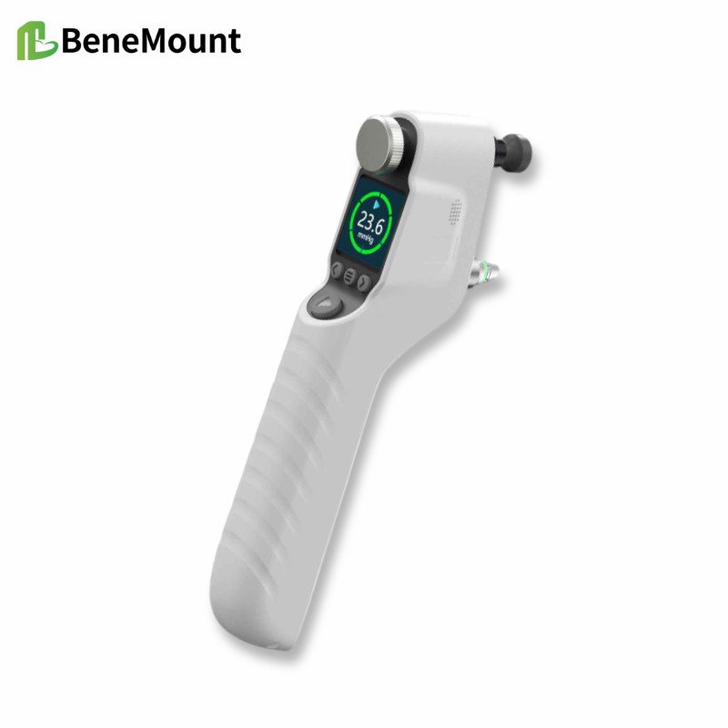
This week’s product is the Eagle Eye V700 veterinary tonometer. The product files describe a rebound veterinary tonometer with animal modes for canine, feline, lapine, equine, primate, rat, and mouse use, and color-coded data quality after repeated measurements. Product information should be used for specifications only; clinical interpretation still requires a complete ophthalmic examination.
References
[1] Gelatt KN, Gilger BC, Kern TJ, eds. Veterinary Ophthalmology. Wiley-Blackwell. Local reference PDF.
[2] Maggs DJ, Miller PE, Ofri R. Slatter's Fundamentals of Veterinary Ophthalmology. Saunders/Elsevier. Local reference PDF.
[3] Gould D, McLellan GJ, eds. BSAVA Manual of Canine and Feline Ophthalmology. BSAVA. Local reference PDF.
[4] Companion Animal Ophthalmology. Local Chinese reference PDF.
[5] Small Animal Ophthalmology. Local Chinese reference PDF.
[6] Kontiola AI. A new induction-based impact method for measuring intraocular pressure. Acta Ophthalmologica Scandinavica. 2000;78(2):142-145.
[7] Knollinger AM, La Croix NC, Barrett PM, Miller PE. Evaluation of a rebound tonometer for measuring intraocular pressure in dogs and horses. Journal of the American Veterinary Medical Association. 2005;227(2):244-248.
[8] Product files for this week: Eagle Eye V700 veterinary tonometer, Chinese manual, quick guide, brochure, English manual, English quick guide, and English brochure. Product files were used only to verify model name, workflow cues, and visible specifications.