

How Veterinary Clinics Can Standardize Pupil Reflex Testing
Why workflow matters more than a single result
PLR testing is easy to perform but easy to perform inconsistently. If each clinician uses a different light, distance, room brightness, exposure time, and wording, the medical record becomes hard to compare.
A standardized workflow does not need to make every case complicated. Its purpose is to make initial exams, rechecks, and referrals comparable. In uveitis, glaucoma, retinal disease, and neuro-ophthalmic cases, serial findings can be more useful than one “normal” or “abnormal” note [1,2].
A practical workflow should define environment, sequence, documentation, and review.
Control the environment first
Use a room where lighting can be controlled. Avoid direct bright light before PLR testing, and allow a short period of dark adaptation when the case allows.
Check recent medications. Tropicamide, atropine, pilocarpine, sedatives, and some systemic drugs can change pupil size or movement. Uveitis, iris atrophy, posterior synechia, corneal edema, and mature cataract can also alter the result.
Restraint should be gentle. Neck pressure, fear, and pain can change autonomic tone. Cats and brachycephalic dogs often require slower handling. A forced test is often a poor test.
A useful clinic sequence
First, observe both eyes from a distance for pupil size, strabismus, eyelid position, opacity, and pain. Second, assess pupil shape and symmetry under normal room light. Third, in dim light, stimulate each eye and record direct and consensual PLR.
Fourth, when useful, apply red and blue light stimuli and observe onset, amplitude, latency, sustained constriction, and escape. Fifth, test menace response, dazzle reflex, obstacle navigation, or cotton ball tracking. Sixth, proceed to slit lamp examination, tonometry, fluorescein staining, fundus examination, and other tests.
This order collects non-contact information before tests that may irritate the eye or alter the pupil. Painful emergencies still require clinical judgment; the workflow should never delay treatment.
How to document the result
A consistent template should include environment, medications, pupil size, right direct and consensual PLR, left direct and consensual PLR, red-light response, blue-light response, dynamic changes, and associated findings.
Example: “Dim room, 2-minute adaptation; no mydriatic used; pupils moderately dilated OU; right direct PLR slow and right-to-left consensual slow; left direct and consensual present; red-light response reduced OD; blue-light response delayed but sustained OD; no obvious pupil escape.”
This is much more useful than “PLR weak.” It tells the next clinician whether the abnormality is more likely afferent, efferent, bilateral, or technique-related.
Recheck and quality control
At recheck, use the same sequence, similar room conditions, and the same light source if possible. Compare speed, amplitude, and maintenance rather than presence alone.
Clinics can train with representative cases: corneal opacity, cataract, glaucoma, retinal disease, and neurologic disease. This helps the team separate media-related artifacts from true pathway abnormalities.
Avoid vague words. Instead of “fair response,” write “constriction begins within 2 seconds, amplitude small, partial redilation after 5 seconds of continued illumination.” Specific records are easier to compare.
Related Product Information
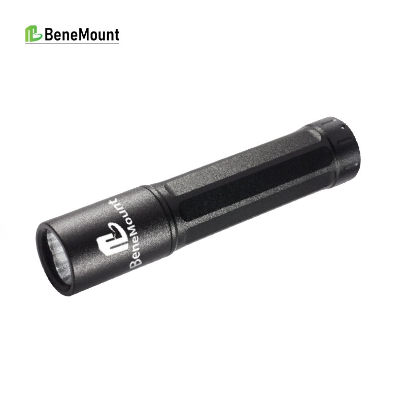
The CPRL Tester can serve as a fixed light source in a standardized clinic workflow. Its value increases when the clinic also standardizes distance, exposure time, red/blue stimulus use, and documentation language.
References / 参考文献
[1] Gelatt KN, Gilger BC, Kern TJ, eds. Veterinary Ophthalmology. 5th ed. Wiley-Blackwell; 2013. Local reference PDF: Kirk N. Gelatt – Veterinary Ophthalmology Two-Volume Set.
[2] Maggs DJ, Miller PE, Ofri R. Slatter's Fundamentals of Veterinary Ophthalmology. 4th ed. Saunders Elsevier; 2008. Local reference PDF.
[3] Gould D, McLellan G, eds. BSAVA Manual of Canine and Feline Ophthalmology. BSAVA; local reference PDF.