

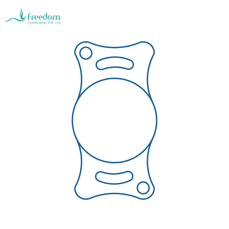
The weakest point in a canine or feline cataract case is not always the operation itself. Problems often arise from an incomplete pathway: late recognition, delayed referral, inadequate preoperative inflammation control, insufficient owner understanding, or interrupted postoperative medication. IOL implantation can improve the optical result in suitable patients, but it cannot replace case management. A pathway from first presentation to recheck is more valuable than a product-centered discussion.[1,2]
The first step is recognition. When a primary veterinarian identifies lens opacity, the record should include visual response, dazzle reflex, intraocular pressure, corneal status, inflammatory signs, and diabetic history. Diabetic dogs, rapidly progressive cataracts, abnormal pressure, nonvisible fundus, or uveitis should prompt early ophthalmic referral. Waiting until a cataract is mature is not always rational, particularly when lens-induced inflammation is likely.[1-4]
The second step is preoperative stabilization. Anti-inflammatory therapy, pressure control, systemic disease management, and owner education determine whether surgery begins with a quiet eye and reasonable capsular conditions. Owners should understand that cataract surgery is not a single transaction. It is a treatment period involving frequent topical medication, protection, and multiple rechecks. If aftercare is unlikely, timing and expectations should be reconsidered.
The third step is intraoperative decision-making. The ideal plan is to implant an appropriately sized canine or feline IOL in an intact capsular bag. If posterior capsule rupture, zonular abnormality, inadequate bag support, or increased inflammatory risk is encountered, ocular safety takes priority. IOL implantation is not a ritual that must be completed; it is a choice that should serve long-term vision and intraocular stability. Experienced teams prepare multiple IOL options and contingency plans.
The fourth step is postoperative monitoring. Rechecks should focus on anterior chamber inflammation, intraocular pressure, corneal clarity, IOL position, posterior capsule clarity, and retinal status. Owner descriptions of vision are useful but cannot replace ophthalmic examination. If an animal again bumps into objects, avoids stairs, or hesitates in low light, the clinician should systematically evaluate PCO, pressure elevation, corneal edema, retinal disease, and IOL displacement.
The pathway should include case review. After each cataract surgery, the team can record cataract stage, diabetic status, preoperative inflammation score, whether an IOL was implanted, implant model, intraoperative complications, postoperative pressure peak, PCO development, and owner feedback. Reviewing these data after three to six months helps the hospital identify which cases need earlier referral, which owners need stronger education, and which surgical steps need improvement.
For a hospital building an ophthalmology service, IOL inventory should support the clinical pathway. Common sizes should be reliably available, high-risk cases should have contingency options, and owner education materials and recheck forms should correspond to implant records. In that setting, a canine or feline intraocular lens is not just a consumable; it is one component of a cataract care system.
Professional educational articles can support this pathway as well. They should help referring veterinarians recognize when to refer, help owners understand why rechecks matter, and help the hospital team use consistent language about cataract surgery and IOL implantation. Product information can appear at the end, but the main body should always solve a clinical problem.
Referring veterinarians also play a key role in the pathway. Even if they do not perform cataract surgery, they can improve prognosis through early recognition, systemic disease control, avoidance of inappropriate long-term medication, and timely referral. In diabetic dogs and cats with suspected uveitis, early ophthalmic assessment is often more valuable than waiting for complete visual loss.
Hospitals can standardize recheck timing, for example day one, week one, week three to four, and then according to inflammation and pressure. If the same indicators are recorded at each visit, the team can identify cases that deviate from normal recovery sooner. IOL implantation is one point in the pathway; continuous management determines program quality.
Related product note: BeneMount lists canine models with 6.00 or 6.50 mm optic options and 11.00 to 15.00 mm overall lengths, and feline models with a 7.00 mm optic and 12.00 to 14.00 mm overall lengths. The material is described as hydrophilic acrylic. A hospital can treat this veterinary IOL as one implant option within a broader cataract pathway and continue to review outcomes through case records.[10]
参考文献
[1] Gelatt KN, Ben-Shlomo G, Gilger BC, Hendrix DVH, Kern TJ, Plummer CE, eds. Veterinary Ophthalmology. 6th ed. Wiley-Blackwell; 2021.
[2] Maggs DJ, Miller PE, Ofri R. Slatter's Fundamentals of Veterinary Ophthalmology. 6th ed. Elsevier; 2018.
[3] Gould D. Clinical assessment of cataracts in dogs. In Practice. 2002;24:46-55.
[4] Appelboam H. Canine cataracts. Companion Animal. 2024.
[5] Kelman CD. Phaco-emulsification and aspiration: a new technique of cataract removal. American Journal of Ophthalmology. 1967.
[6] Awasthi N, Guo S, Wagner BJ. Posterior capsular opacification: a problem reduced but not yet eradicated. Archives of Ophthalmology. 2009;127(4):555-562.
[7] Nibourg LM, Gelens E, Kuijer R, Hooymans JMM, van Kooten TG, Koopmans SA. Prevention of posterior capsular opacification. Experimental Eye Research. 2015;136:100-115.
[8] Findl O, Buehl W, Bauer P, Sycha T. Interventions for preventing posterior capsule opacification. Cochrane Database of Systematic Reviews.
[9] American Academy of Ophthalmology. Cataract in the Adult Eye Preferred Practice Pattern. 2021.
[10] BeneMount. 适应各种需求的犬猫用人工晶体产品彩页. 上海仁山医疗器械有限公司.
产品资料摘录