

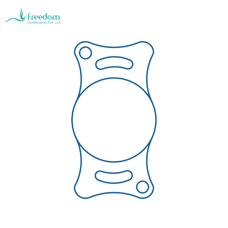
IOL material is not an isolated product attribute; it becomes a clinical variable during surgery. One advantage of phacoemulsification is that lens removal can be performed through a smaller incision. If a rigid IOL requires the incision to be enlarged, part of the minimally invasive benefit is lost. The value of a foldable intraocular lens is that implantation can remain consistent with the small-incision logic of phacoemulsification.[5,9]
Hydrophilic acrylic IOLs generally provide flexibility and foldability, allowing insertion through an injector or folding system into the anterior chamber and capsular bag. For the veterinary surgeon, the relevant observations are practical: loading stability, controlled entry, smooth unfolding, need for manipulation, and capsular stress. The intraocular space in dogs and cats is limited; the corneal endothelium, iris, and posterior capsule should not be exposed to repeated attempts. Material behavior matters when it reduces tissue disturbance and intraocular handling time.[1,2]
Aspheric design relates to image quality. A spherical optic can contribute to spherical aberration, whereas an aspheric design aims to optimize light focus and contrast. Animals do not read acuity charts, but functional vision is still important. Can the dog judge stair height? Can the cat move confidently in low light? Can the patient track a toy or owner gesture? Clinical communication should present aspheric optics as an image-quality design rather than as a vague premium feature.[1,2]
Material also interacts with inflammation and long-term clarity, but the relationship should not be oversimplified. PCO reviews indicate that material, edge design, capsular bag contact, and inflammation all contribute to posterior capsule change. In dogs and cats, preoperative uveitis, diabetes, owner compliance, and follow-up frequency may be as important as material differences. A good surgical team does not treat material as insurance against complications; it integrates material choice into perioperative strategy.[6,7]
When evaluating a veterinary IOL, hospitals can record model, lot, delivery method, main-incision size, unfolding time, need for repositioning, anterior chamber reaction at day one and week one, intraocular pressure, corneal clarity, and IOL centration. After a meaningful number of cases, the team can judge whether the material and design fit its surgical workflow and patient population.
Foldable IOL implantation also requires team coordination. The surgical nurse should understand lens handling, hydration, loading orientation, and injector preparation. The surgeon must coordinate OVD protection, incision size, capsular opening, and implantation speed. Many difficulties arise not from the implant itself but from unfamiliarity with the delivery sequence. When a new product is introduced, dry-lab rehearsal and workflow confirmation are often safer than first use in a complex case.
Aspheric design and optic size should also be considered in relation to pupil behavior. Canine and feline pupil size is strongly influenced by medication, pain, inflammation, and light. Postoperative pupil distortion may magnify optic-edge issues. If iris atrophy, synechiae, or pupil deformation is present, the surgeon should consider whether IOL centration and optic coverage will be adequate. Material and optical design are most useful when intraocular structures remain reasonably stable.
Material selection should not be separated from sterility, packaging, and traceability. An IOL is an implant. Lot number, expiration date, sterilization status, and storage conditions should be part of the surgical checklist. If these details are recorded, later inflammation, clarity, or positioning concerns can be investigated through meaningful quality tracking.
If a hospital introduces a new canine or feline IOL, it is wise to begin with routine cases that have favorable intraocular conditions rather than complex trauma, severe uveitis, or zonular instability. Material and delivery systems have a learning curve. The team should first confirm loading, insertion, unfolding, and documentation workflows before using the implant in more complex eyes.
Material evaluation should include surgeon handling impression, but it should not rely on feel alone. A lens that loads smoothly and unfolds predictably is valuable, but subjective experience should be paired with objective findings: whether the incision needed enlargement, whether the anterior chamber remained stable, whether the cornea was clear postoperatively, whether the IOL centered well, and whether inflammation was controlled. Experience becomes reliable when it is recorded.