• Home Main
  • All Products
    • Bandage lenses
    • Biologic grafts
    • Diagnostic Tools
    • ERG/ cPLR
    • Eye Surgery Tools
    • Fundoscopy
    • Ophthalmic Suture
    • Phaco Device and Accessories
    • Slit lamp
    • Surgical instruments
    • Tonometer
    • Shipping Fee
    • Ophthalmic Anatomy Charts
  • ECM
  • About us
  • Blog
  • Contact us
  • Warranty Policy
Search
0 items $0.00
Login / Register
Menu
0 items $0.00
Home All Products Ophthalmic Anatomy Charts Anatomical Diagram of the Feine Eye
BeneMount®SLp-05-Lab Veterinary Tabletop Slit Lamp Laboratory Kit
BeneMount®SLp-05-Lab Veterinary Tabletop Slit Lamp Laboratory Kit $6,500.00
Back to products
BeneMount Universal Adjustable Laboratory Rodents Bracket —— BeneMount®SLp-labC
BeneMount Universal Adjustable Laboratory Rodents Bracket —— BeneMount®SLp-labC $850.00
Anatomical Diagram of the Canine Eye
Click to enlarge

Anatomical Diagram of the Feine Eye


You save

✅ Clinical Anatomy & Pathology: Connects key ocular structures with common clinical signs and eye conditions, supporting clearer explanations during veterinary consultations.

✅ Client Education & Communication: Helps veterinarians explain complex eye anatomy, diagnoses, and procedures through detailed visual illustrations.

✅ Practical Training Reference: Ideal for veterinary interns, technicians, and nursing staff. A2 landscape format with anti-glare matte lamination for everyday clinical use.

Contact us

This product is currently out of stock and unavailable.

Email when stock available

Import duties and taxes: Import duties, taxes, and customs clearance fees, if applicable, are not included in the product price or shipping fee and must be paid by the recipient.
Compare
  • Description
  • Additional information
  • QR Code
Description

BENEMOUNT · VETERINARY EDUCATION

Make Eye Anatomy Easier to Explain

Canine & Feline Ophthalmic Anatomy Posters bring detailed illustrations into everyday veterinary practice. A practical visual reference for client consultations, clinical discussions, and team training.

A2 landscape   /   Anti-glare matte finish   /   English or Chinese
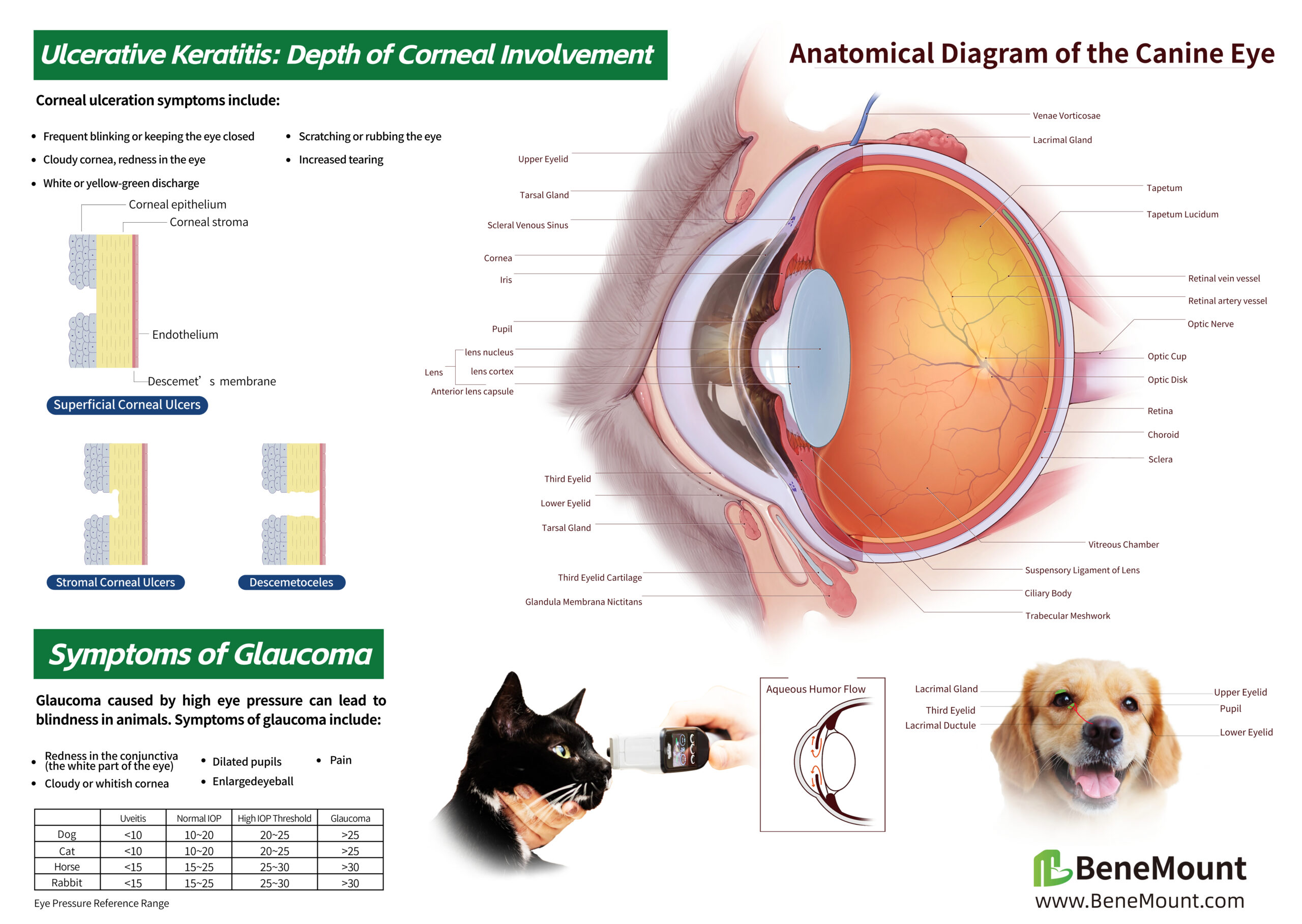
Canine eye anatomy poster with labeled ocular structures, corneal ulcer diagrams and glaucoma information

01 / CANINE EYE ANATOMY

From the Whole Eye to the Clinical Conversation

Explain the cornea, anterior chamber, lens, retina, and optic nerve with a detailed, labeled cross-section of the canine eye.

Supporting illustrations connect ocular anatomy with corneal ulceration, aqueous humor flow, and common signs associated with glaucoma, giving consultations a clear visual starting point.

For the consultation room: Point to the structure, explain the finding, and help owners follow the discussion.

Enlarged detail from the canine poster comparing superficial corneal ulcers, stromal corneal ulcers and descemetoceles

02 / CORNEAL ULCER DETAIL

Show Why Ulcer Depth Matters

A closer look at the canine poster highlights three illustrated patterns: superficial corneal ulcers, stromal corneal ulcers, and descemetoceles.

The side-by-side cross-sections make the depth of corneal involvement easier to visualize, supporting explanations of examination findings, monitoring, and recommended care.

Useful for team training: Refer to the same illustration during case discussions and owner education.
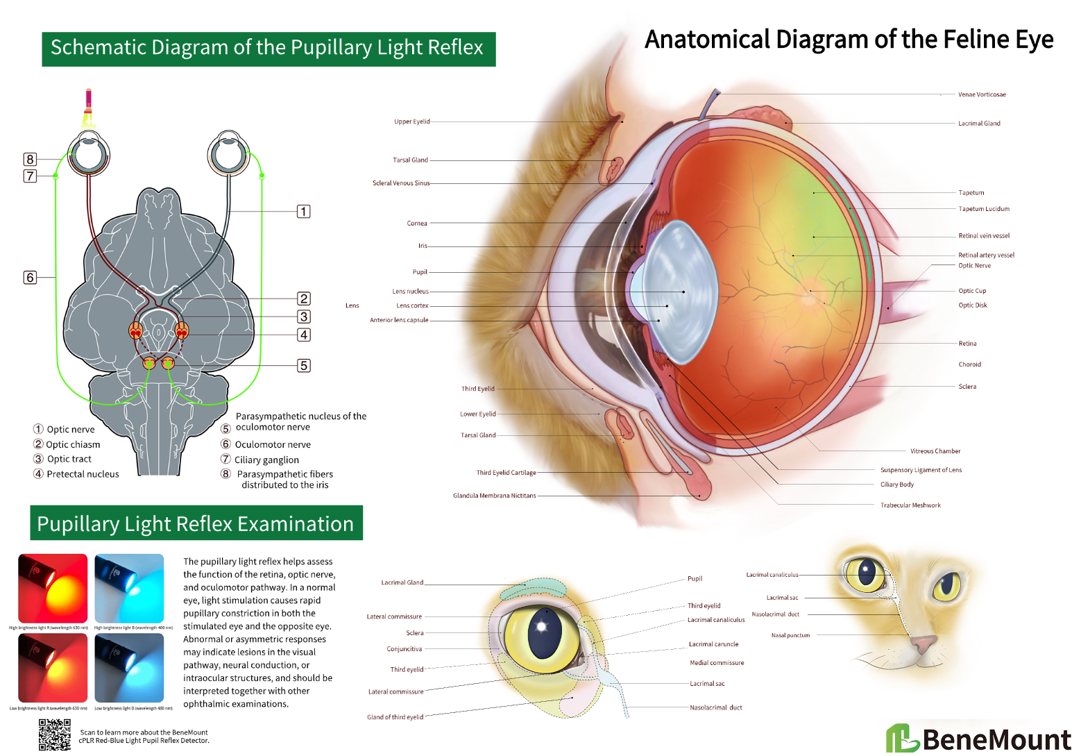
English feline eye anatomy poster with labeled cross-section, pupillary light reflex pathway and tear drainage anatomy

03 / FELINE EYE ANATOMY

A Detailed View of the Feline Eye

Bring feline ocular anatomy into focus with a labeled cross-section, external eye landmarks, and illustrations of the lacrimal system.

The poster combines eye structure with a schematic of the pupillary light reflex, helping teams connect anatomical landmarks with the examination process.

For client education: Use clear visual references to explain the eye, eyelids, and tear drainage during consultations.

Enlarged feline pupillary light reflex pathway with optic nerve, optic chiasm, optic tract, pretectal nucleus, oculomotor nerve and ciliary ganglion labels

04 / PUPILLARY LIGHT REFLEX DETAIL

Follow the Pathway Behind the Reflex

The enlarged schematic traces the pupillary light reflex through the optic nerve, optic chiasm, optic tract, and pretectal nuclei, then follows the parasympathetic pathway to the iris.

Color-coded pathways and numbered labels provide a shared visual reference for discussing direct and consensual pupillary responses during team training.

From structure to examination: Keep the pathway visible while explaining how light stimulation leads to pupillary constriction.

MADE FOR EVERYDAY PRACTICE

A Clear Reference, Ready When You Need It

Support veterinary interns, technicians, and nursing staff with an accessible visual reference for in-house training and daily clinical discussions. Matte lamination helps reduce reflections from examination lights and overhead lighting.

Product Specifications

  • Size: A2 — 594 × 420 mm / 23.4 × 16.5 in
  • Orientation: Landscape
  • Finish: Anti-glare matte lamination
  • Species options: Canine / Feline
  • Languages: English / Chinese

Choose Your Version

  • Canine Eye Anatomy — English
  • Canine Eye Anatomy — Chinese
  • Feline Eye Anatomy — English
  • Feline Eye Anatomy — Chinese

IN YOUR PRACTICE

See It in Your Consultation Room

See how each A2 eye anatomy poster fits into a veterinary consultation room, keeping a clear visual reference close at hand.
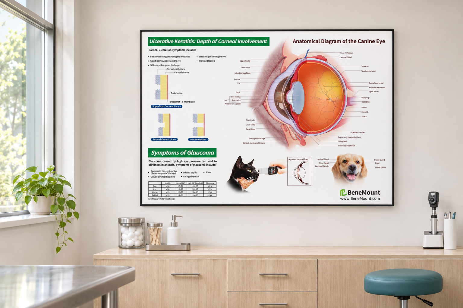
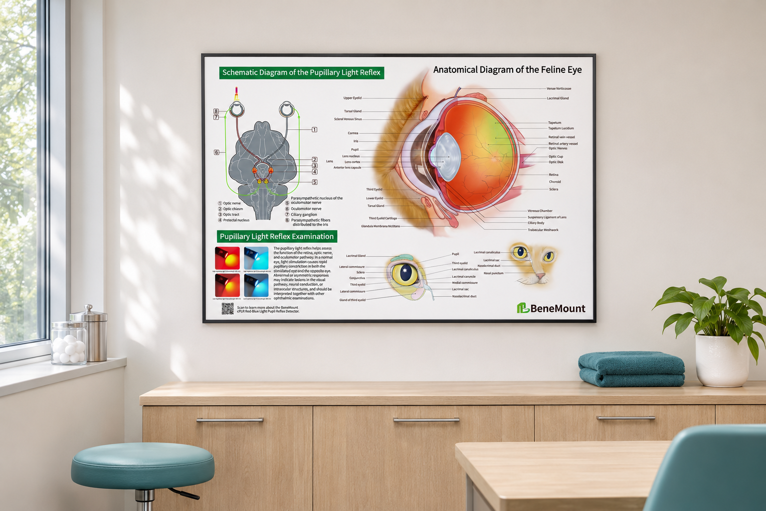
BeneMount canine eye anatomy poster displayed individually on a veterinary consultation room wall
Canine Eye Anatomy
BeneMount feline eye anatomy poster displayed individually on a veterinary consultation room wall
Feline Eye Anatomy

Simulated displays for illustration. Frames and room furnishings are shown for styling purposes.

Additional information
Species & Language

Canine – English, Canine – Chinese, Feline – English, Feline – Chinese

QR Code
QR Code

SKU: N/A Category: Ophthalmic Anatomy Charts Tag: price_basic
Share:

Related products

Compare
Quick view

BeneMount Corneal Trephine – Short

In stock

$18.00
You save
Select options
SMI Ophthalmic Sutures Absorbable
Compare
Quick view

SMI Ophthalmic Sutures Absorbable

In stock

$99.00 – $119.00Price range: $99.00 through $119.00
You save
Select options
Opticlear Two Mirror Goniolenses
Compare
Quick view

Opticlear Two Mirror Goniolenses

In stock

$150.00
Add to cart
Compare
Quick view

BeneMount Conjunctival Scissors ​Curved(105mm)

In stock

$30.00 – $45.00Price range: $30.00 through $45.00
You save
Select options
Rose Bengal Test Strips
Compare
Quick view

Rose Bengal Strips

In stock

$22.00
Add to cart
Compare
Quick view

BeneMount Corneal Scissors​ Curved(116mm)

In stock

$30.00 – $45.00Price range: $30.00 through $45.00
You save
Select options
​Ophthalmic Silicone Sterilization Case
Compare
Quick view

​Ophthalmic Silicone Sterilization Case

In stock

$22.00 – $56.00Price range: $22.00 through $56.00
You save
Select options
Reichert® Ocu-Film® + Tip Covers
Compare
Quick view

Reichert® Ocu-Film® + Tip Covers (Box of 150, individually wrapped)

In stock

$145.00
Add to cart
Useful Links
  • About us
  • Shop
  • Contact us
Legal
  • Terms of Service
  • Shipping Policy
  • Refund and Returns Policy
  • Privacy Policy
  • Warranty Policy
Contact Us

  • info@wisevisionintl.com
  • RM 522-523,5/F INTERNATIONAL PLAZA NO 20 SHEUNG YUET RD KOWLOON BAY KLN HONG KONG

Copyright © 2024 Wise Vision International Co.,Limited All Rights Reserved.
  • Menu
  • Categories
  • Home Main
  • All Products
    • Bandage lenses
    • Biologic grafts
    • Diagnostic Tools
    • ERG/ cPLR
    • Eye Surgery Tools
    • Fundoscopy
    • Ophthalmic Suture
    • Phaco Device and Accessories
    • Slit lamp
    • Surgical instruments
    • Tonometer
    • Shipping Fee
    • Ophthalmic Anatomy Charts
  • ECM
  • About us
  • Blog
  • Contact us
  • Warranty Policy
  • Home Main
  • All Products
    • Bandage lenses
    • Biologic grafts
    • Diagnostic Tools
    • ERG/ cPLR
    • Eye Surgery Tools
    • Fundoscopy
    • Ophthalmic Suture
    • Phaco Device and Accessories
    • Slit lamp
    • Surgical instruments
    • Tonometer
    • Shipping Fee
    • Ophthalmic Anatomy Charts
  • ECM
  • About us
  • Blog
  • Contact us
  • Warranty Policy
  • Compare
  • Login / Register
Shopping cart
Close
Sign in
Close

Lost your password?

No account yet?

Create an Account
Anatomical Diagram of the Canine Eye

Anatomical Diagram of the Feine Eye


You save
Select options
Compare
Start typing to see products you are looking for.
Shop
0 items Cart
My account