


BENEMOUNT · VETERINARY EDUCATION
Make Eye Anatomy Easier to Explain
Canine & Feline Ophthalmic Anatomy Posters bring detailed illustrations into everyday veterinary practice. A practical visual reference for client consultations, clinical discussions, and team training.
A2 landscape / Anti-glare matte finish / English or Chinese
01 / CANINE EYE ANATOMY
From the Whole Eye to the Clinical Conversation
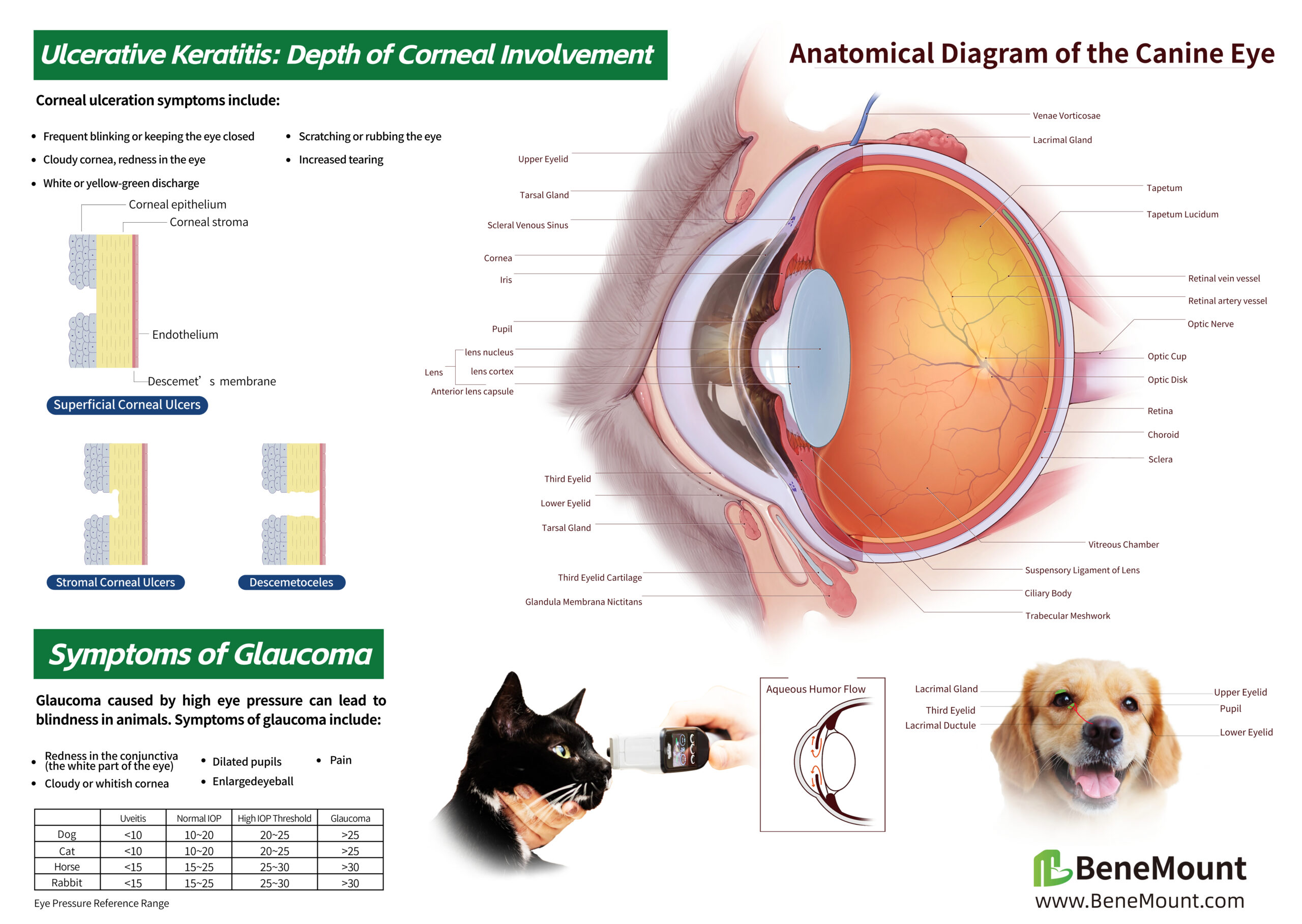
Explain the cornea, anterior chamber, lens, retina, and optic nerve with a detailed, labeled cross-section of the canine eye.
Supporting illustrations connect ocular anatomy with corneal ulceration, aqueous humor flow, and common signs associated with glaucoma, giving consultations a clear visual starting point.
For the consultation room: Point to the structure, explain the finding, and help owners follow the discussion.
02 / CORNEAL ULCER DETAIL
Show Why Ulcer Depth Matters
A closer look at the canine poster highlights three illustrated patterns: superficial corneal ulcers, stromal corneal ulcers, and descemetoceles.
The side-by-side cross-sections make the depth of corneal involvement easier to visualize, supporting explanations of examination findings, monitoring, and recommended care.
Useful for team training: Refer to the same illustration during case discussions and owner education.
03 / FELINE EYE ANATOMY
A Detailed View of the Feline Eye
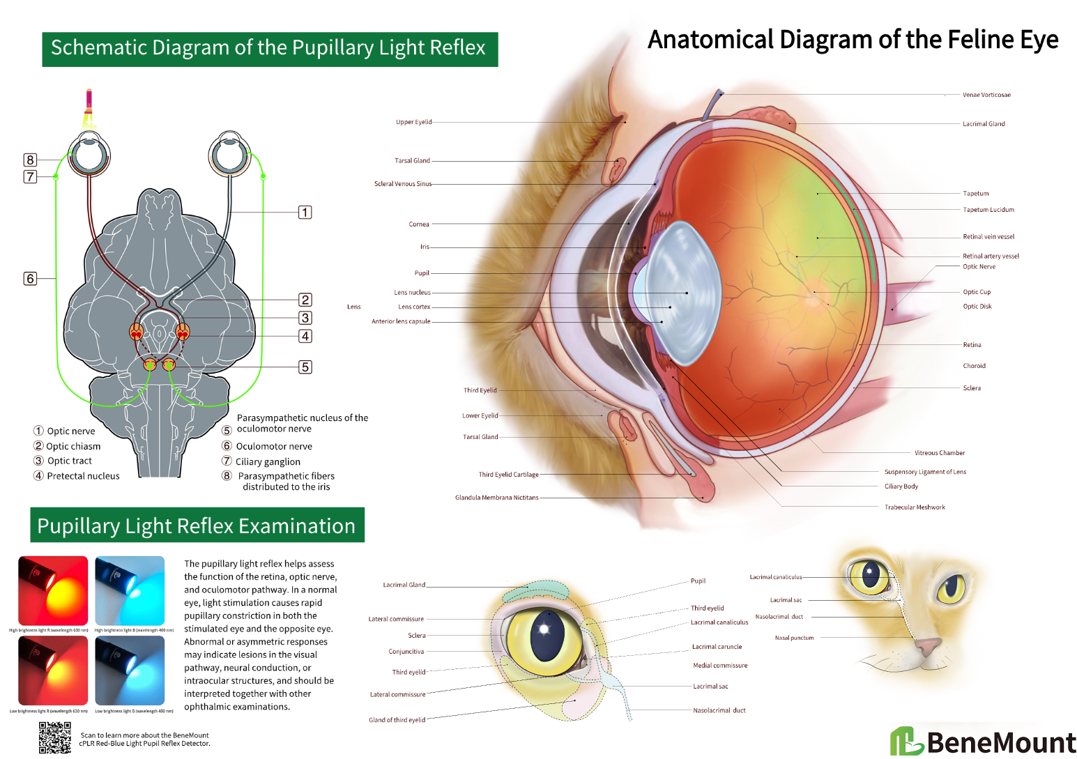
Bring feline ocular anatomy into focus with a labeled cross-section, external eye landmarks, and illustrations of the lacrimal system.
The poster combines eye structure with a schematic of the pupillary light reflex, helping teams connect anatomical landmarks with the examination process.
For client education: Use clear visual references to explain the eye, eyelids, and tear drainage during consultations.
04 / PUPILLARY LIGHT REFLEX DETAIL
Follow the Pathway Behind the Reflex
The enlarged schematic traces the pupillary light reflex through the optic nerve, optic chiasm, optic tract, and pretectal nuclei, then follows the parasympathetic pathway to the iris.
Color-coded pathways and numbered labels provide a shared visual reference for discussing direct and consensual pupillary responses during team training.
From structure to examination: Keep the pathway visible while explaining how light stimulation leads to pupillary constriction.
MADE FOR EVERYDAY PRACTICE
A Clear Reference, Ready When You Need It
Support veterinary interns, technicians, and nursing staff with an accessible visual reference for in-house training and daily clinical discussions. Matte lamination helps reduce reflections from examination lights and overhead lighting.
Product Specifications
- Size: A2 — 594 × 420 mm / 23.4 × 16.5 in
- Orientation: Landscape
- Finish: Anti-glare matte lamination
- Species options: Canine / Feline
- Languages: English / Chinese
Choose Your Version
- Canine Eye Anatomy — English
- Canine Eye Anatomy — Chinese
- Feline Eye Anatomy — English
- Feline Eye Anatomy — Chinese
IN YOUR PRACTICE
See It in Your Consultation Room
See how each A2 eye anatomy poster fits into a veterinary consultation room, keeping a clear visual reference close at hand.
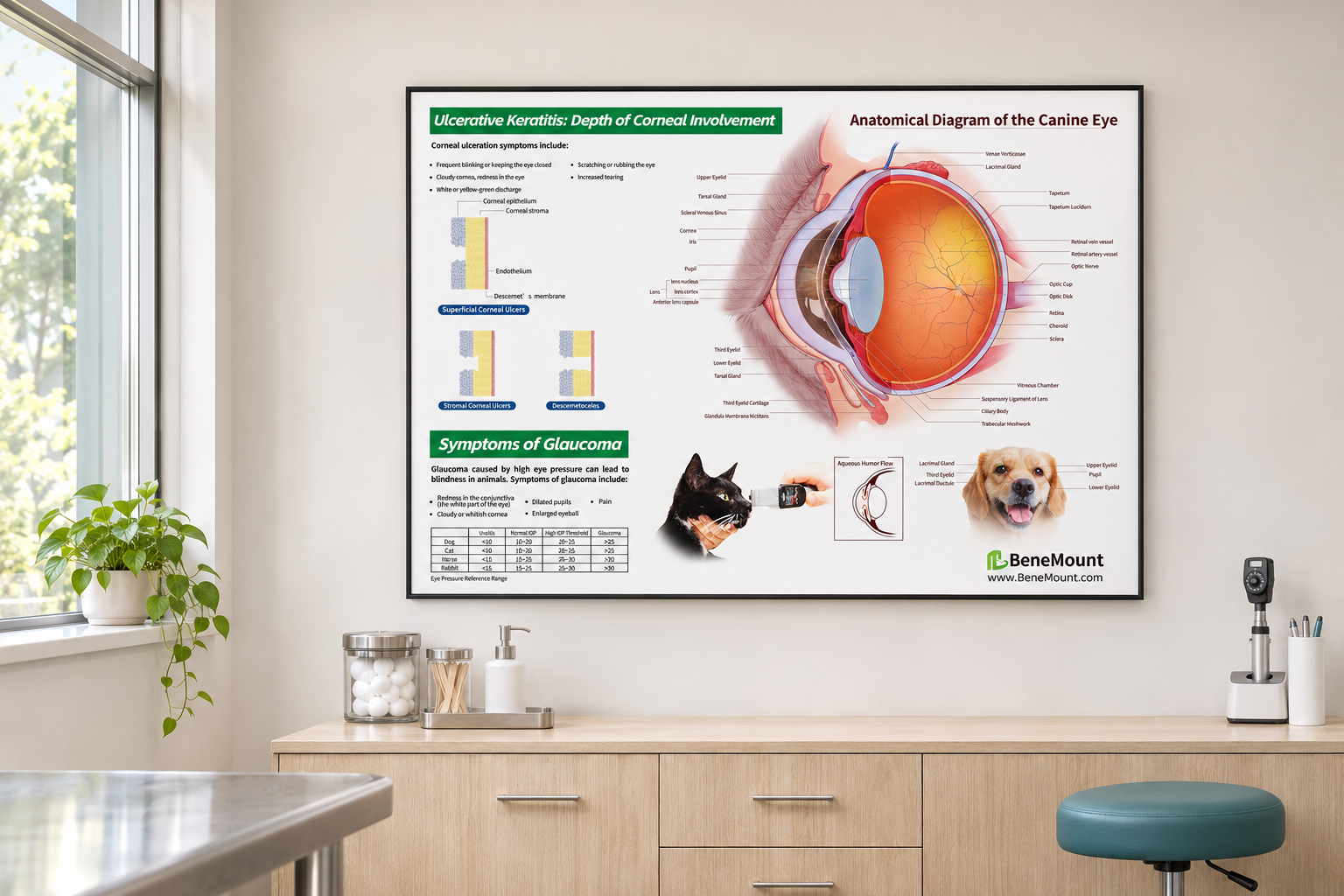
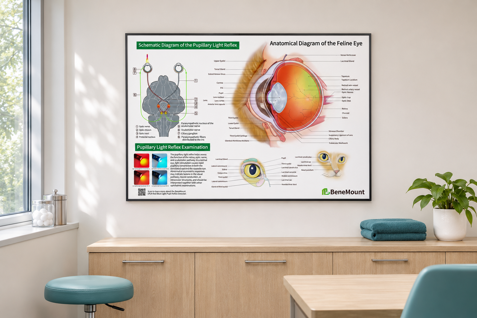
Simulated displays for illustration. Frames and room furnishings are shown for styling purposes.
