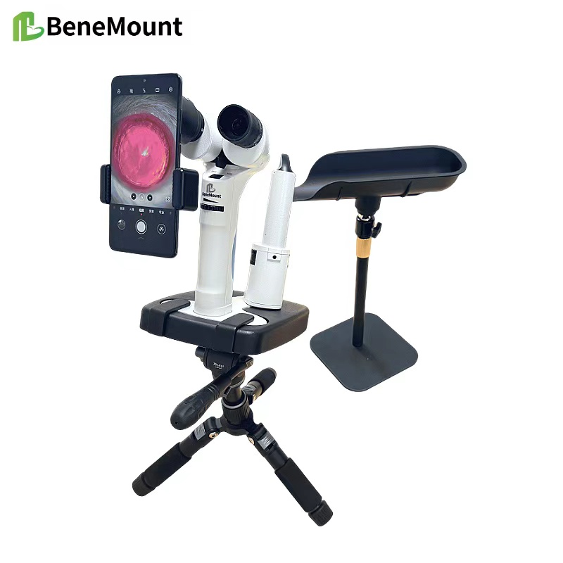
Slit-Lamp Biomicroscopy Training is now a key component of modern veterinary education. For students, mastering this technique provides not only technical competence but also a deeper understanding of ocular anatomy and disease. By integrating structured instruction with clinical exposure, educators can prepare future veterinarians to use the slit lamp effectively in both small animal and large animal practice.
Teaching the Fundamentals
The foundation of training begins with familiarization. Students must first learn how to set up the instrument, adjust magnification, and manipulate the light beam. Classroom sessions often include demonstrations of diffuse, direct, and retroillumination. Early lessons emphasize hand–eye coordination and the importance of maintaining patient comfort during examination. By practicing on models or cooperative animals, students build confidence before moving to clinical cases.
Developing Examination Skills
Once basic handling is secure, the focus shifts to structured eye exams. Students learn to identify corneal layers, anterior chamber depth, iris architecture, and lens clarity. Educators encourage systematic routines: starting with diffuse overview, then narrowing to optical sections, and finally applying retroillumination for fine changes. Repetition strengthens both technical skill and clinical judgment, ensuring students can recognize subtle abnormalities in real patients.
Clinical Integration
Practical rotations give students the opportunity to apply slit-lamp biomicroscopy on actual veterinary cases. Under supervision, they examine dogs with corneal ulcers, cats with uveitis, and horses with early cataracts. This exposure highlights the diagnostic value of the slit lamp and builds problem-solving ability. Regular feedback from instructors allows refinement of technique, while case discussions link clinical findings to pathology and treatment decisions.
Overcoming Challenges
Student training also addresses common difficulties. Hand tremor, incorrect beam alignment, or overuse of bright illumination can reduce accuracy and stress patients. Teachers stress the importance of gentle handling and calm positioning. Emphasis is placed on repeatability—students are taught to document findings consistently so that disease progression can be monitored across time.
Conclusion
Effective Slit-Lamp Biomicroscopy Training equips veterinary students with essential diagnostic skills for their future careers. By combining technical instruction, supervised practice, and structured clinical exposure, educators ensure that graduates can use this instrument with confidence. Mastery of slit-lamp examination enhances diagnostic accuracy, supports early detection of disease, and strengthens the overall standard of veterinary ophthalmology.


