

The Role of Tonometry in the Diagnosis and Management of Glaucoma in Animals
Glaucoma is a group of chronic progressive diseases characterized by persistently elevated intraocular pressure (IOP), ultimately resulting in optic nerve atrophy and irreversible loss of visual function. In clinical veterinary practice, glaucoma is particularly prevalent in dogs, where it is one of the most common causes of blindness. The disease can also occur in cats, horses, rabbits, and other species, though less frequently.
The Role of Tonometry in the Diagnosis and Management of Glaucoma in Animals
Etiology and Types
Animal glaucoma can be classified into primary and secondary forms.
Primary glaucoma is mostly hereditary, frequently seen in certain dog breeds such as Basset Hounds, Cocker Spaniels, Shih Tzus, and Beagles. The pathogenesis of primary glaucoma mainly involves structural abnormalities of the iridocorneal angle, resulting in impaired aqueous humor outflow.
Secondary glaucoma arises from other ocular diseases—such as uveitis, lens luxation, neoplasia, trauma, or hyphema—which impede aqueous humor drainage. Secondary glaucoma is more common in species such as cats and horses; for instance, cats often develop glaucoma due to chronic uveitis causing angle closure.
Pathogenesis
The core pathological change in glaucoma is obstruction of aqueous outflow leading to elevated IOP.
Under normal circumstances, aqueous humor is produced by the ciliary body and exits via the trabecular meshwork and scleral venous channels. When these outflow pathways are blocked, aqueous accumulates within the eye, resulting in increased IOP. Sustained high IOP directly damages the nerve fibers of the optic nerve head, destroys retinal ganglion cells, and ultimately leads to irreversible vision loss. At the same time, elevated IOP can also injure other intraocular structures, including the cornea, lens, and retina, resulting in complications such as corneal edema, hyphema, and secondary cataracts.
Clinical Manifestations
The clinical signs of animal glaucoma vary depending on the disease course and the rate of progression. Common findings include:
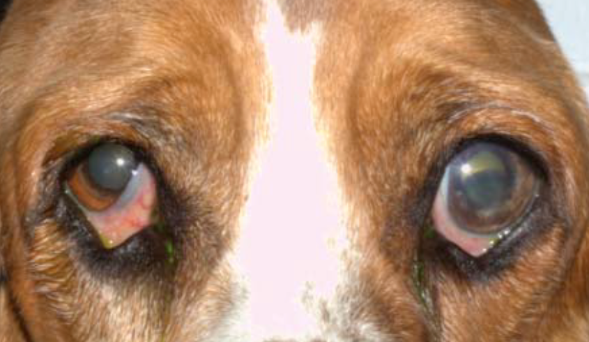
Exophthalmos (buphthalmos, especially in dogs)
Corneal edema (appearing as a blue or cloudy eye surface)
Moderately dilated pupil with sluggish pupillary light response
Marked elevation of IOP (commonly >25–30 mmHg in dogs, >25 mmHg in cats during acute episodes)
Progressive vision loss, possibly complete blindness
Pale and atrophic optic disc in late stages
Signs of discomfort in some animals, such as pain, tearing, or blepharospasm
Occasionally, anterior displacement of the globe, shallow anterior chamber, iris color changes, or vitreous haze
Susceptible Species and Breeds
Glaucoma is most commonly seen in dogs, with pronounced breed predispositions (such as American Cocker Spaniels, Basset Hounds, Akitas, and Poodles). In cats, glaucoma is mostly secondary, especially in elderly cats with chronic uveitis or synechiae of the iridocorneal angle. In horses, glaucoma is often associated with recurrent uveitis; in rabbits, it is rare and mostly related to congenital anomalies. There are also reports of glaucoma in some wild animals (e.g., lions, giraffes, hippos) and laboratory species.
Prognosis and Outcome
The prognosis of glaucoma is closely linked to the timeliness of diagnosis and treatment.
If acute high IOP is not controlled within several hours to days, irreversible damage to the retina and optic nerve frequently leads to complete blindness in the short term, and most dogs will develop phthisis bulbi or globe loss within months. Chronic or secondary glaucoma progresses more slowly, but long-term vision preservation is still limited.
In most cases, medical or surgical interventions can only delay progression and relieve pain; restoration of lost vision is extremely rare. Therefore, early and accurate monitoring and intervention of IOP are key to delaying the course toward blindness.
Identifying Early Unilateral Disease: The Value of Bilateral Comparison
In many breeds with primary angle-closure glaucoma, the disease often progresses in a stepwise manner—first affecting one eye, then the other. The textbook emphasizes:
The difference in IOP between both eyes should be less than 8 mmHg; if this threshold is exceeded, both eyes should be carefully examined for potential pathology.
In clinical practice, if one eye’s IOP is 18 mmHg (normal), while the other reaches 28–30 mmHg, even in the absence of overt pain or corneal edema, one must be alert to subacute glaucoma or developmental angle abnormalities. Bilateral comparison also helps exclude systemic factors (e.g., blood pressure, sedatives, body position) that could cause generalized IOP fluctuations, thus more precisely identifying the affected eye. For species prone to secondary glaucoma, such as cats and horses, this is equally important: one eye may develop elevated IOP due to uveitis-induced synechiae, while the other remains in a compensatory phase. Early detection and timely intervention can significantly delay vision loss in the contralateral eye.
Quantitative Indices for Staging, Prognosis, and Dynamic Monitoring
Precisely because the pathogenesis of glaucoma is highly dependent on abnormal elevation of IOP, and the speed of disease progression and prognosis are closely related to the level of IOP, only scientific and standardized tonometry allows early detection, timely intervention, and dynamic follow-up. Tonometry not only provides veterinarians with quantitative and objective data, but also enables clinicians to accurately determine the disease stage, evaluate treatment efficacy, and anticipate disease changes. Thus, tonometry is essential in all stages of glaucoma—from screening and diagnosis to long-term management—its importance in veterinary ophthalmology cannot be overstated.
Quantification and Tracking
Each IOP measurement yields a specific value and coefficient of variation. Repeated IOP measurements are crucial for disease monitoring and treatment evaluation. Electronic medical records allow visualization of IOP trends in the same animal across different dates and times, aiding assessment of therapeutic effects or disease progression.
Consistency Across Devices and Operators
Modern electronic tonometers (such as Tono-Vera Vet) are equipped with self-test and calibration modules, which control variables such as probe pressure, contact time, and angle deviation. This ensures a high degree of comparability, even when different operators or clinics perform the measurements. Studies show that experienced operators can reproduce results within ±5 mmHg 100% of the time in canine eye models, and well-trained novices can also achieve over 90% repeatability.
The Role of Tonometry in the Diagnosis and Management of Glaucoma in Animals
Clear Clinical Decision Thresholds
The textbook provides clear warning values: ≥25 mmHg in dogs and cats, an interocular difference >8 mmHg, or persistent IOP ≥30 mmHg all require urgent intervention. Objective values can rapidly trigger clinical workflows for “medication adjustment—reassessment—surgical consultation.”
Thanks to its objectivity, quantification, and repeatability, tonometry is recognized as the “gold standard” for long-term management of glaucoma in animals.
References
All data and quotations are from:
Kirk N. Gelatt – Veterinary Ophthalmology Two-Volume Set-Wiley-Blackwell, Section II: Foundations of Clinical Ophthalmology, pp. 621–629, Ch. 20, Table 10.1.5.