Introduction
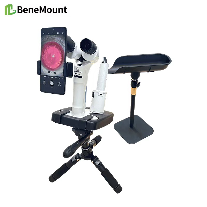
Slit-Lamp Biomicroscopy is central to the clinical evaluation of dogs and cats. It allows veterinarians to examine the anterior eye with precision and magnification. In everyday practice, the technique helps to detect disease early, guide treatment, and monitor response over time. The ability to visualize corneal and lenticular changes in detail makes the slit lamp a critical tool for small animal ophthalmology.
Corneal Disease Assessment
In dogs and cats, the cornea is a frequent site of pathology. Slit-lamp examination helps the clinician assess epithelial defects, stromal edema, and endothelial integrity. A narrow optical section reveals the depth of ulcers, while diffuse illumination shows the extent of opacity. Retroillumination can highlight subtle scars or keratic precipitates on the posterior cornea. Accurate corneal assessment informs both medical and surgical planning.
Evaluation of the Anterior Chamber
The anterior chamber provides key information about ocular health. Inflammatory cells, flare, or hyphema become visible when illuminated at an oblique angle. Early anterior uveitis, which may be missed in routine inspection, becomes clear under the slit beam. Measurement of anterior chamber depth also aids in identifying lens luxation or predisposition to glaucoma.
Iris and Pupil Examination
Changes in iris texture and color are best documented by slit-lamp inspection. Nodules, atrophy, or neoplastic infiltration can be recognized at an early stage. The pupillary margin and sphincter function can also be assessed with focused illumination. In cats, detection of subtle iris melanoma often depends on careful slit-lamp study.
Lens and Cataract Detection
Early lens opacities are difficult to see without magnification. Slit-Lamp Biomicroscopy reveals the location and density of cataracts, allowing staging and prognosis. Nuclear sclerosis, incipient cortical cataracts, or lens capsule ruptures can be differentiated with precision. Such findings guide both medical management and referral for surgery.
Clinical Value in Routine Practice
For both dogs and cats, slit-lamp biomicroscopy bridges the gap between gross inspection and advanced imaging. It is a repeatable and objective method that improves diagnostic confidence. Regular use enhances early disease detection, which is critical for preserving sight and ensuring animal welfare.
Conclusion
The slit lamp is indispensable for small animal ophthalmology. By mastering illumination techniques and patient handling, veterinarians can unlock its full diagnostic potential. The greatest strength of slit-lamp biomicroscopy lies in its ability to reveal ocular changes at a stage when treatment can still prevent irreversible vision loss.


