

Learn about the Schirmer Tear Test (STT) in veterinary ophthalmology. Understand how to perform and accurately interpret STT results for animal eye health.
What is the Schirmer Tear Test
The pre-corneal tear film consists of three layers. The outer layer is the lipid layer, produced by the meibomian glands; the middle layer is the aqueous layer, produced by the lacrimal gland and the gland of the third eyelid; the inner layer is the mucin layer, originating from conjunctival goblet cells.
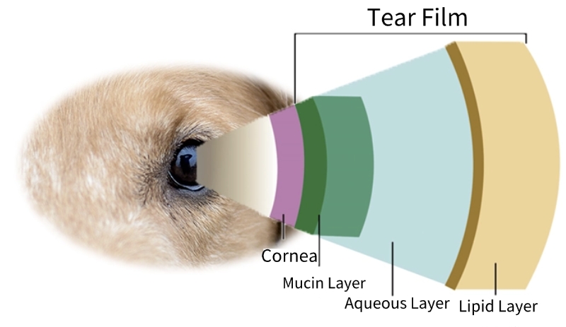
The animal Schirmer Tear Test (STT) is the primary diagnostic method for Keratoconjunctivitis Sicca (KCS) (or dry eye disease). It assesses the aqueous layer of the tear film, and results are reported in millimeters of wetting on the test strip per minute.
By combining the tear production volume with clinical signs, it can be determined whether the animal has KCS and the severity of the condition.
Types of Schirmer Tear Test
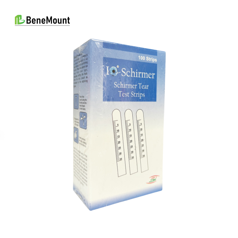
Schirmer Tear Test Strips
The Schirmer Tear Test (STT) is typically divided into Type I (STT I) and Type II (STT II). Both types of tear tests use the same test strips but differ in method.
STT I does not require topical anesthesia and is primarily used to evaluate both the animal’s basal and reflex tear secretion; it is widely applied in small animal clinical practice.
STT II requires the use of a topical anesthetic and is mainly used to evaluate the animal’s basal tear secretion; it is not widely applied in small animal clinical practice.
Indications
When is a Schirmer Tear Test (STT) indicated for animals?
- Symptoms of Ocular Dryness: If an animal exhibits symptoms such as dry eyes, pain, itching, redness and swelling, increased discharge, or general ocular discomfort, performing an STT is recommended to assess tear volume and determine the presence of dry eye disease (KCS) or other ocular conditions.
- Ocular Disease Diagnosis: When diagnosing certain ocular conditions, STT can be part of the overall ocular health assessment. This includes, for example, corneal ulcers, conjunctivitis, keratitis, and eyelid abnormalities.
- Ocular Trauma Assessment: After an animal experiences ocular trauma or injury, STT can be used to assess the eye’s tear production capacity to determine if ocular dryness or other underlying issues exist.
- Pre-surgical Assessment: In cases requiring ocular surgery, STT can be part of the preoperative evaluation. This helps ascertain the eye’s pre-surgical health status and tear production capacity.
- Medication Monitoring: Certain drug therapies can affect an animal’s tear production. STT can be used to monitor changes in tear production during treatment and to determine the effectiveness of the therapy.
- Screening of Breeds Predisposed to KCS: Screening animal breeds prone to KCS is crucial for early diagnosis and management, helping to provide timely treatment and protect the animal’s ocular health. Breeds susceptible to KCS include: English Bulldog, Shih Tzu, West Highland White Terrier, Pekingese, American Cocker Spaniel, Boston Terrier, Miniature Schnauzer, Samoyed, Poodle, Bichon Frise, and others.
- Geriatric Ocular Disease Screening: As animals age, ocular health issues become more common, including conditions like KCS, cataracts, glaucoma, and other eye diseases.
The above are some common situations where STT may be indicated. However, in veterinary clinical practice, the decision of whether an animal should undergo STT ultimately rests with the veterinarian based on the animal’s specific condition.
STT I Examination Procedure and Precautions
1. Preparation for Examination
Schirmer Tear Test Strips
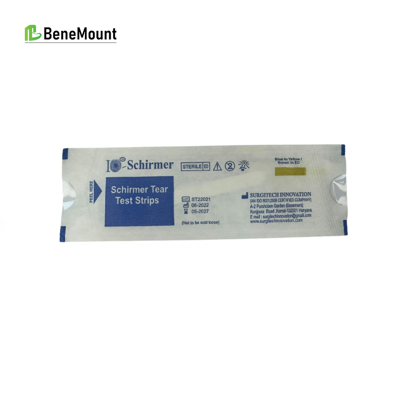
Schirmer Tear Test Strips
Surgical Timer
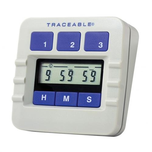
Surgical Timer
2. Steps for STT
1) Animal restraint: Ensure safety while applying minimal necessary restraint.
Dog: Place one hand behind the head, and the other hand under the chin.
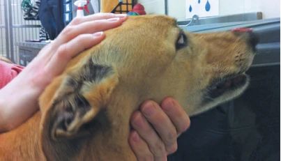
schirmer tear test dogs
Cat: Place two thumbs behind the ears and your index fingers under the chin.
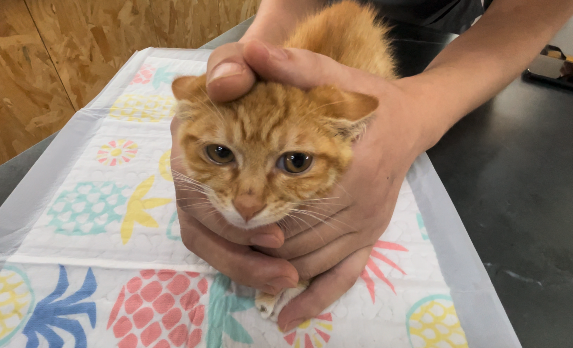
schirmer tear test cats
2) Fold the rounded end of the test strip inside the sterile packaging, thus maintaining the sterility of the strip’s measuring end.
◼︎ Caution: Do not touch the measuring end of the Schirmer test strip, as skin oils can affect the results.
3) Remove the test strip from its packaging.
Insert the bent, rounded tip of the strip between the animal’s lower eyelid and the cornea, approximately one-half to two-thirds of the way from the medial canthus along the length of the eyelid.
◼︎ The eye being tested can be either closed or open. If the animal is unwilling to remain still, you can gently close its eyelids for assistance.
4) After the test strip has been in place for 1 minute, immediately remove it and record the reading on the strip. The result is the value indicated by the meniscus line at the junction of the dry and wet areas on the strip.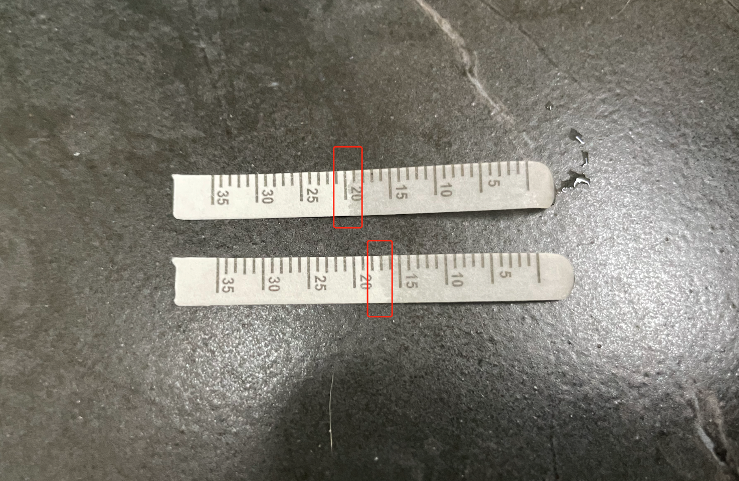
◼︎ Regardless of whether the test strip has a color developing agent, the measurement result should be based on the wet-dry demarcation line of the test strip.
3. Important Notes
- STT I (Schirmer Tear Test I) should be performed before other irritating or ocular medication examinations to prevent errors in test results.
- During STT I, avoid excessive pressure on the eyelids and keep the animal’s eyes away from direct light.
- This test must be performed before applying any other liquid to the eye.
- If thick mucus is present in the animal’s cornea or conjunctival sac, it should be gently removed with a cotton ball or gauze before measurement to prevent interference with the test.
STT I Result Interpretation
STT results must be interpreted in conjunction with the animal’s other clinical signs.
1.Reference Range
Canine:
If a dog’s STT I value is below 15 mm/min, Keratoconjunctivitis Sicca (KCS) can be suspected. In actual clinical practice, canine STT readings can generally be interpreted as follows:
- ≥ 15 mm/min = Normal range
- 11-14 mm/min = Early KCS
- 6-10 mm/min = Moderate KCS
- ≤ 5 mm/min = Severe KCS
Feline:
KCS (Keratoconjunctivitis Sicca) is rare in cats. A cat’s tear glands are controlled by the sympathetic nervous system, and during examination, a cat might experience sympathetic nervous system arousal due to stress, leading to reduced tear secretion. In actual clinical practice, feline STT readings can generally be interpreted as follows:
- ≥ 7 mm/min = Normal range
2.Factors Leading to Result Deviations
1) Pathological Increase (↑)
- Eyelash Disorders (e.g., Distichiasis/Ectopic Cilia): Ingrown or ectopic eyelashes can irritate the eye and stimulate increased tear production.
- Corneal Diseases: Certain corneal conditions, such as ulcers or inflammation, can stimulate excessive tear secretion.
- Uveitis: Ocular inflammation caused by uveitis may lead to an increase in tear secretion.
- Glaucoma: Glaucoma can cause elevated intraocular pressure, stimulating increased tear secretion.
- Meibomian Gland Dysfunction (MGD): MGD can lead to reduced normal tear film stability and a compensatory increase in aqueous tear production.
- Treatment of Spontaneous Chronic Corneal Epithelial Defects (SCCEDs): Certain treatments for SCCEDs might stimulate increased tear secretion.
- Lacrimal Duct Obstruction: Obstruction or blockage of the tear drainage system prevents normal tear outflow, leading to prolonged tear retention on the ocular surface, which may affect STT results.
2) Pathological Decrease (↓)
- Immune-Mediated Dry Eye (KCS): Immune-mediated dry eye disease can lead to reduced tear production.
- Medications (e.g., Atropine): Certain medications, such as atropine, may inhibit tear secretion, leading to a decrease.
- Upper Eyelid Injury Involving Lacrimal Gland Damage/Inflammation: Upper eyelid injuries can affect the normal function of the lacrimal gland, leading to reduced tear secretion.
- Surgical Excision of the Third Eyelid Gland: If the third eyelid gland is surgically removed, it may result in decreased tear production.
- Viral Infections: Certain viral infections (e.g., Feline Infectious Peritonitis Virus) may affect lacrimal gland function, leading to reduced tear production.
- Neurogenic: Certain neurogenic diseases (e.g., facial nerve paralysis) may lead to reduced tear secretion.
3) Other Influencing Factors (↑↓)
- Environmental Factors: Factors such as humidity, temperature, and airflow in the testing environment can influence tear production. Overly dry or cold environments may lead to insufficient tear production, while overly humid or hot environments might affect the accuracy of the test results.
- Animal’s State: The animal’s emotional state, anxiety, or stress levels can impact tear production. Stressed or anxious animals may produce less tear fluid, leading to lower test results.
- Improper Technique: The operational technique during the testing process can also affect the results. If the filter paper strip is placed incorrectly, excessive pressure is applied, or other procedural errors occur, it can lead to inaccurate results.
- Medication Interference: The use of certain medications or eye drops may interfere with tear production.
To obtain more accurate results, these influencing factors should be minimized as much as possible when performing the STT. This includes conducting the test under appropriate environmental conditions, ensuring the animal’s comfort and relaxation, following correct operating techniques, and, when possible, avoiding medications that might affect tear production.
Most importantly, STT results should always be interpreted in conjunction with the animal’s clinical signs, other ocular examination findings, and medical history.
Disclaimer:
This article is for the purpose of learning and exchanging knowledge in animal ophthalmology only, and does not constitute diagnostic or treatment advice.