
Principles and Clinical Significance of Tonometry in Veterinary Ophthalmology
Introduction
Tonometry, the quantitative measurement of intraocular pressure (IOP), is fundamentally grounded in ocular biomechanics and the hydrodynamics of aqueous humor. Therefore, an accurate determination of IOP is critical. It is essential for understanding eye physiology. Tissue integrity is also revealed. Numerous ophthalmic disorders are illuminated.
The Biophysical Foundations of Tonometry
The eye functions as a partially deformable sphere. It is filled with incompressible fluid. Consequently, intraocular pressure is maintained. This occurs through a dynamic equilibrium. Aqueous humor is secreted by the ciliary processes. Its drainage then happens. This is via trabecular and uveoscleral outflow pathways. A baseline tension within the globe results from this equilibrium. This tension is distributed across the collagenous framework. Both the sclera and cornea are involved. The IOP, therefore, provides structural support for the globe. Furthermore, a crucial reference state is established. This is for the proper function of intraocular tissues.
From a physical perspective, tonometry is the transduction of internal pressure. This is converted into an external measurement. This is accomplished by controlled deformation. The corneal surface is targeted. Subsequent quantification of resistance follows. The response of the cornea and sclera to external force is determined. Their viscoelastic properties are key. These properties are known to vary. Differences exist between species and individuals. Variations also occur under different pathological conditions.
Measurement Principles and Evolution of Techniques
Indentation Tonometry
Indentation tonometry is the earliest method. Historically, it was widely employed. In this technique, the depth of corneal indentation is measured. This is caused by a known weight. The Schiøtz tonometer is a classic example. A weighted plunger is placed on the anesthetized cornea. The extent of indentation can then be measured. This is done on a linear scale. It is then converted to mmHg after calibration. The underlying assumption is important. The eye approximates a thin-walled sphere. The force needed to indent the cornea inversely reflects internal pressure. However, this method is significantly affected. Variations in ocular rigidity pose a challenge. Corneal thickness also impacts results. Scleral elasticity is another factor. Thus, its accuracy is less reliable. This is particularly true in veterinary patients. Diverse globe anatomy is often present.
Applanation Tonometry
Applanation tonometry builds upon the Imbert-Fick principle, which states that the pressure inside an ideal sphere equals the force required to flatten a known area of its surface divided by that area. In practical terms, instruments like the Tono-Pen and Goldmann-type tonometers apply a small, flat-tipped probe to the cornea and measure the force necessary to applanate a fixed area. The cornea’s capillary attraction and rigidity are accounted for in instrument design and calibration. Electronic applanation tonometers, such as the Mackay–Marg or Tono-Pen, provide digital readouts and minimize observer bias. These instruments are less influenced by variations in ocular rigidity compared to indentation tonometry, though corneal thickness, curvature, and even the tear film can still affect measurements.
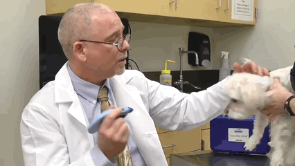
Tonometry in Veterinary Ophthalmology-Applanation Tonometry
Rebound Tonometry
Rebound tonometry represents a more recent innovation. A small probe is propelled toward the cornea and the deceleration of the probe as it contacts and rebounds is analyzed. The deceleration characteristics correlate with the intraocular pressure: higher IOP results in more rapid deceleration and shorter contact time. This method, typified by the Tonovera-Vet, requires no topical anesthesia and is particularly well suited for small eyes, exotics, and patients in which restraint is challenging. Rebound tonometry is less affected by corneal pathology or thickness and offers improved applicability across species, but care must be taken to standardize probe orientation and measurement location.
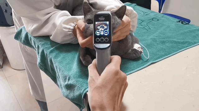
Tonometry in Veterinary Ophthalmology-Rebound Tonometry
Technical Influences and Potential Sources of Error
The accuracy of tonometry is not absolute. All techniques make assumptions about the physical and anatomical properties of the eye. Deviations from these assumptions—such as changes in corneal thickness, local scarring, edema, or variations in scleral rigidity—will alter the relationship between measured force and true intraocular pressure. Operator technique, including the positioning of the animal, avoidance of digital or jugular pressure, and consistent measurement site, is also critical. Moreover, pharmacological agents, especially mydriatics, sedatives, and anesthetics, can alter IOP and thus must be considered when interpreting results.
Direct manometry, involving direct cannulation and measurement of pressure within the anterior chamber, is considered the reference standard, but its invasiveness restricts its use to experimental or intraoperative settings. For most clinical scenarios, the combination of appropriate technique, understanding of the chosen instrument’s calibration, and awareness of patient-specific variables are required to ensure both accuracy and repeatability of IOP measurement.
Conclusion
The principles of tonometry are deeply rooted in ocular biomechanics and physics. Modern veterinary ophthalmology benefits from a spectrum of tonometric instruments, each based on a unique measurement principle and each with inherent strengths and limitations. A comprehensive understanding of these underlying principles enables clinicians to select appropriate methods, interpret findings with nuance, and contribute to the highest standard of ocular diagnosis and management.