Introduction
Animal Slit-Lamp Examination is an essential procedure in veterinary ophthalmology. It allows clinicians to evaluate the anterior eye with magnification and controlled illumination. Reliable results depend not only on equipment quality but also on examiner technique and patient management. By understanding key precautions, veterinarians can reduce errors and improve diagnostic accuracy.
Preparing the Instrument
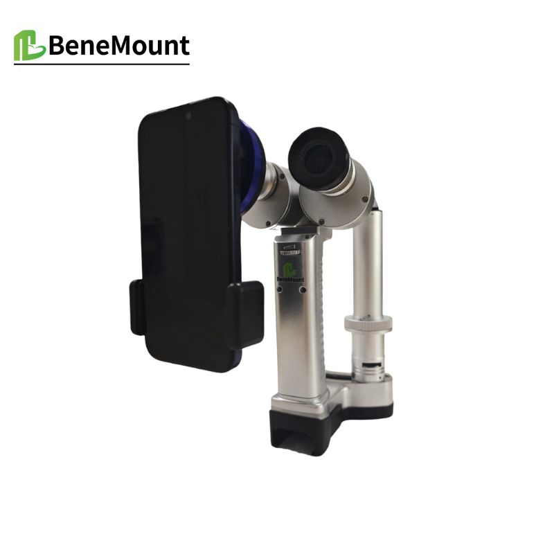
Accurate examination begins with proper preparation of the slit lamp. Clean optics are vital, since dust or smudges can mimic ocular pathology. Illumination should be adjusted to a level tolerated by the patient. The clinician should start with low magnification to orient the view, and only then increase for detailed inspection. Before every session, chin or head rests must be disinfected to protect both patient and staff.
Patient Handling and Positioning
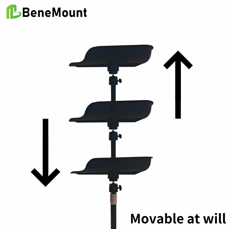
Animals do not remain still as human patients do, so careful restraint is necessary. Dogs and cats are usually examined on the table, with gentle manual support to keep the head steady. Any pressure on the neck or periocular tissues must be avoided, since this can raise intraocular pressure and distort findings. Horses may require portable slit lamps or mild sedation to achieve cooperation. Small mammals and birds often demand upright positioning, which reflects natural physiology. Comfort and stability are the guiding principles across species.
Illumination Techniques and Their Limits
A wide beam provides a general survey, but too much light can startle cats or rabbits. Narrow beams reveal optical sections of cornea and lens, yet precise alignment is required. Direct focal illumination highlights lesions on the ocular surface, while retroillumination uncovers subtle opacities by reflecting light from deeper structures. Each method has its limits: glare, reflex tearing, or sudden movement can alter interpretation. The examiner must adapt technique in real time, maintaining both accuracy and patient tolerance.
Factors That Compromise Accuracy
Several common mistakes can reduce the reliability of an Animal Slit-Lamp Examination. Improper restraint, bright illumination in a photophobic animal, or misalignment of the beam may all produce misleading images. Inconsistent positioning between sessions can also hinder longitudinal comparison. Environmental factors such as poor room lighting, noise, or stress from handling further complicate the procedure. Recognizing these pitfalls allows clinicians to anticipate and minimize them.
Clinical Significance
When performed with care, slit-lamp biomicroscopy provides diagnostic details impossible to obtain by gross inspection. Subtle uveitis, early corneal dystrophy, and small lens opacities can be identified before they affect vision. Regular, standardized use also ensures that treatment outcomes can be monitored objectively. In this way, slit-lamp examination is not only a diagnostic tool but also a cornerstone of long-term ocular health management.
Conclusion
Precision in veterinary ophthalmology depends on both technology and technique. By observing clear precautions in setup, handling, and illumination, the clinician can ensure reliable results. The true clinical value of an Animal Slit-Lamp Examination lies in its capacity to provide early, accurate insight into diseases that may otherwise remain undetected.