

Precautions and Common Pitfalls in Animal Tonometry
Patient Handling and Positioning
First, always handle patients gently and minimize restraint. When you apply excessive pressure to the neck, head, or periocular area, you risk artificially increasing IOP. For this reason, never hold the animal tightly around the neck or press on the globe. Instead, support the head with your hand placed beneath the jaw or behind the ears, depending on the species.
Body and head position also matter. Ensure that the animal is comfortably positioned, ideally with the head at or slightly above heart level. In horses and some other large animals, lowering the head below heart level can cause a noticeable elevation in IOP. In small mammals and birds, holding the animal upright usually provides the most physiologically accurate measurement.
Animal Tonometry
Environmental and Behavioral Factors
Reducing patient stress is another crucial step. Anxiety, struggling, and vocalization may all temporarily raise IOP, especially in cats and rabbits. Therefore, prepare a quiet environment and, whenever possible, allow the animal to acclimate before examination. For fractious or highly anxious animals, you may need to reschedule tonometry or consider mild sedation, always noting the use of sedatives in your records.
Use of Topical Anesthesia
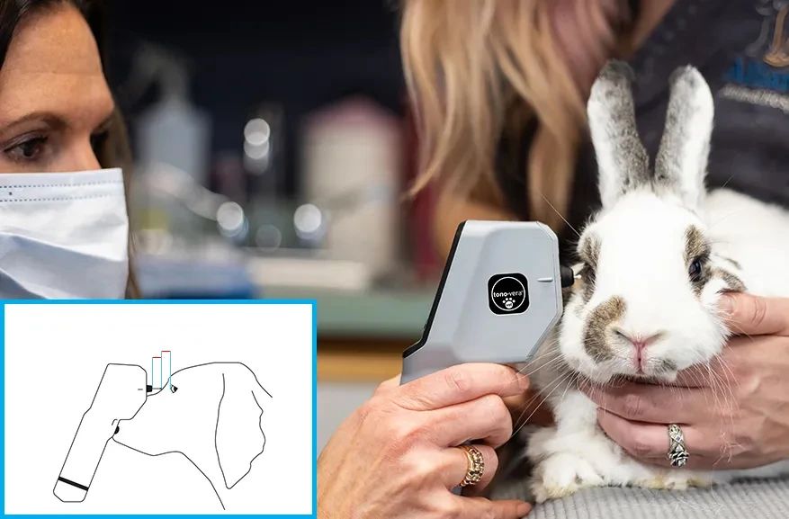
When using applanation tonometers such as the Tonopen Avia Vet, always apply topical anesthetic before measurement. Inadequate anesthesia can result in increased blinking or movement, which affects both accuracy and patient comfort. Conversely, for rebound tonometry with devices such as the Tono-Vera Vet, topical anesthesia is usually unnecessary; however, if the animal shows signs of discomfort or has corneal disease, you can apply anesthetic as needed.
Device Handling and Calibration
Proper device preparation ensures consistent results. Always calibrate the tonometer according to the manufacturer’s instructions before each session. For devices with disposable tips or probes, install a new sterile cover for every patient to prevent cross-contamination and measurement errors.
When you position the tonometer, always keep the probe perpendicular to the corneal surface and avoid tilting the instrument. Repeatedly tapping the same corneal area can cause surface changes or artifactually alter the pressure reading. Therefore, if you need to repeat the measurement, move to a slightly different location on the central cornea.
Avoiding Operator-Induced Artifacts
Common operator-induced errors include applying pressure to the eyelids or globe during measurement. Even a small amount of extra pressure may significantly increase the IOP reading, leading to a false diagnosis of ocular hypertension or glaucoma. Similarly, using a tight collar or holding the neck can obstruct venous return and raise IOP, particularly in brachycephalic dogs.
Furthermore, avoid taking measurements immediately after a prolonged struggle, restraint, or recent application of mydriatic or anesthetic drugs unless absolutely necessary. These interventions can alter IOP for several minutes to hours.
Interpreting Results: Bilateral and Serial Measurements
Always compare the IOP between both eyes, as a difference greater than 8 mmHg is generally considered abnormal and may signal unilateral pathology. However, do not rely solely on a single “snapshot” value. Repeat the measurement at different times of day and monitor changes over time, especially when managing chronic conditions such as glaucoma.
Influence of Corneal Disease and Ocular Surface
Corneal pathology—including edema, ulcers, scarring, or irregularity—can interfere with tonometry accuracy. For example, measurements taken over an area of corneal edema may be falsely high or low, depending on the tonometer type. Whenever possible, choose the most normal-appearing area of the cornea for measurement and document any abnormalities.
In addition, excessive tearing, ocular discharge, or the presence of topical medications (especially viscous ointments) can also affect the contact between probe and cornea. Clean and dry the ocular surface gently before proceeding.
Device-Specific Pitfalls
For the Tonopen Avia Vet, always use a new Ocu-Film tip for each animal, and be sure to verify that the device is properly calibrated before every session. With the Tono-Vera Vet, regularly check probe alignment and use the targeting system for consistent, perpendicular contact. In both cases, failure to follow manufacturer instructions for maintenance and calibration may result in systematically inaccurate readings.
Documentation and Communication
Finally, carefully document every measurement, including the animal’s species, age, clinical status, restraint method, use of anesthetic or sedatives, device settings, and any challenges encountered during the session. Sharing these details with colleagues ensures continuity of care and helps avoid misinterpretation of isolated results.