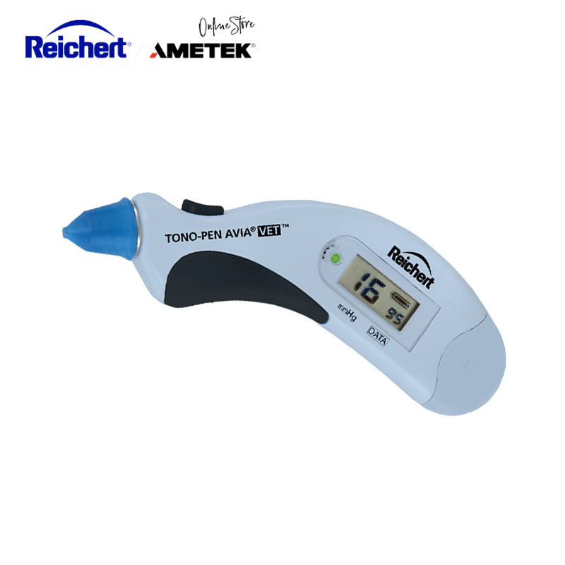
Influence of Sedation, Anesthesia, and Body Position on Tonometry Readings in Animals
In clinical veterinary ophthalmology, the influence of sedatives, anesthetic agents, and body position on intraocular pressure (IOP) measurement is often underestimated, but these are actually major non-pathological factors that can lead to falsely elevated or reduced readings. Understanding these influences and adhering to standardized protocols is essential for obtaining reliable diagnostic data and avoiding misdiagnosis or overtreatment.
Effects of Sedative Agents
Different classes of sedatives affect animal IOP through distinct mechanisms and with varying degrees of impact.
Alpha-2 adrenergic agonists (xylazine, detomidine, medetomidine) generally lower IOP. Xylazine can reduce IOP by 2–7 mmHg in dogs and horses by decreasing aqueous humor production, inducing vasoconstriction, and reducing stress-induced spikes.
Benzodiazepines (diazepam, midazolam) have little effect on IOP in dogs and cats, though deep sedation with respiratory depression may slightly increase IOP. Phenothiazines (acepromazine) have minimal effect and are suitable when stability is desired.
Dissociative anesthetics (ketamine, tiletamine-zolazepam) significantly increase IOP in dogs and cats (8–15 mmHg), related to increased BP and extraocular muscle tone. Not recommended when accurate readings are critical.
Effects of Anesthetic Agents
The effect of general anesthetics varies by agent, dose, route, and species.
Inhalational anesthetics (isoflurane, sevoflurane) usually lower IOP by vasodilation and reduced muscle tone. However, deep anesthesia with hypoxemia or hypercapnia can cause unpredictable rises.
Barbiturates and propofol lower IOP effectively, but excessive dosing may cause CO₂ retention, transiently raising IOP.
Opioids (morphine, fentanyl) have little effect and can control pain without altering IOP significantly.
Effect of Body Position and Cervical Pressure
Body and head position strongly influence readings.
- Dogs & Cats: Sternal recumbency is most stable. Dorsal or head-down positions reduce IOP. Any cervical pressure or jugular compression can raise IOP by 10–20 mmHg or more.
- Horses: IOP rises if the head is below heart level. Keep the head level or slightly elevated.
- Small mammals & birds: Upright or natural standing reflects physiological IOP best.
Clinical Recommendations and Practical Points
- Avoid tonometry under sedation/anesthesia unless necessary (esp. in suspected glaucoma).
- If drugs are required, measure baseline IOP first and record drug, dose, and timing.
- Keep the head level with or above the heart; avoid cervical restraint.
- Take multiple readings and average; always compare both eyes.
- Document all influencing factors for later interpretation.


