
How to Use Veterinary Tonometers: Step-by-Step Guide for Common Species
Introduction
Accurately measuring intraocular pressure (IOP) is essential in veterinary ophthalmology for both diagnosis and ongoing management of ocular diseases. With advancements in technology, modern veterinary tonometers like the Reichert Tono-Vera Vet and Tonopen Avia Vet allow veterinarians to quickly and reliably assess IOP across a wide range of animal species. Knowing how to use these instruments effectively, while adapting techniques for different patients, is crucial for obtaining clinically meaningful results.
Preparation and Patient Handling
Before beginning tonometry, you should create a calm and quiet environment to minimize stress and movement in the patient. When handling the animal, always support the head gently and avoid any pressure on the neck or around the eyes. This is especially important for brachycephalic breeds and small mammals, as even mild restraint can lead to artificially elevated IOP readings. Good lighting is essential for visualizing the cornea and properly positioning the veterinary tonometers.
When using the Tonopen Avia Vet or Tono-Vera Vet, topical ocular anesthesia is recommended. Instill a drop of proparacaine or tetracaine in the conjunctival sac and wait one to two minutes for full effect. Anesthesia not only makes the procedure more comfortable for the patient but also ensures more consistent and accurate results.
Step-by-Step Operation:
Tonopen Avia Vet (Applanation Tonometry)
Device Preparation and Calibration
Start by installing a fresh, sterile Ocu-Film tip cover on the Tonopen Avia Vet sensor. Power on the instrument and allow it to complete its self-check. Confirm “Ready” status on the display before proceeding.
Topical Anesthesia
Instill one drop of topical ocular anesthetic (e.g., proparacaine) onto the cornea. Wait about one to two minutes for full effect, which helps minimize patient discomfort and ensures more consistent readings.
Patient Positioning and Restraint
Gently restrain the animal, supporting the head but avoiding any pressure on the neck or around the eyes. Position the patient so that you have a clear, direct view of the corneal surface.
Eyelid Retraction
Use your non-dominant hand to gently open the eyelids, taking care not to touch the globe.
Taking the Measurement
Hold the Tonopen perpendicular to the corneal surface. Tap the tip lightly and briefly against the central or paracentral cornea. The Tonopen collects a series of readings automatically, then calculates and displays the mean IOP and the coefficient of variation. If the coefficient is high (often above 5–10%), repeat the procedure.
Documentation
Record the mean IOP, which eye was tested, the animal’s species, and any notes about the animal’s behavior or ocular surface.
Species Adaptation:
Tonopen Avia Vet is highly versatile and can be used across species—including dogs, cats, rabbits, guinea pigs, birds, reptiles, and horses—making it especially valuable for exotics. Always apply topical anesthetic before use in all species, including small mammals and birds.
Tono-Vera Vet (Rebound Tonometry)
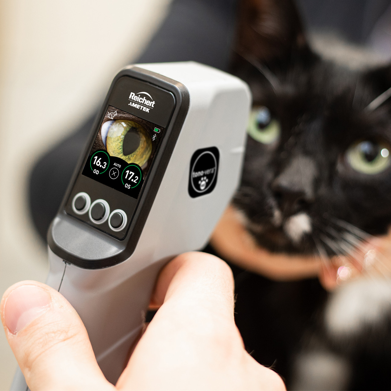
Veterinary Tonometers(Rebound Tonometry)
Device Preparation and Probe Installation
Turn on the Tono-Vera Vet and insert a clean, disposable probe according to the manufacturer’s instructions. The device performs an automatic self-check and displays when it is ready.
Species Selection (if available)
Choose the correct species setting if your device supports this feature (dog, cat, horse, etc.), as the algorithm compensates for corneal biomechanics.
No Topical Anesthesia Needed
In most cases, you do not need to use topical anesthesia, as the rebound probe’s light touch is well-tolerated. However, in extremely sensitive animals or if there is significant corneal disease, anesthesia may be considered.
Patient Positioning and Handling
Gently restrain the animal, minimizing physical restraint and avoiding any neck or ocular pressure. Position the patient so that the eye is facing forward and at your eye level.
Taking the Measurement
Hold the Tono-Vera Vet so that the probe is perpendicular and at the recommended distance (usually 4–8 mm) from the central cornea. Activate the device, allowing the probe to bounce quickly and gently off the corneal surface. The instrument will automatically record several measurements and display the average IOP with a quality indicator.
Quality Control and Repeat Measurements
If the device signals poor alignment or inconsistent results, reposition and repeat the measurement. Tono-Vera Vet’s on-screen targeting (if available) helps you achieve precise alignment.
Documentation
Record the displayed IOP value, which eye was tested, and any notable factors about the session.Using Tonopen Avia Vet and Tono-Vera Vet in Different Species
Dogs and Cats
Both the Tonopen Avia Vet and Tono-Vera Vet perform reliably in dogs and cats. Always avoid compressing the neck or jugular veins, as this can lead to falsely high results. In cats, gentle handling and minimal restraint are especially important, since stress alone may transiently increase IOP. An auriculopalpebral nerve block is not required, but topical anesthesia is standard.
Rabbits, Small Mammals, and Exotics
The Tonopen Avia Vet is particularly well suited for rabbits, ferrets, guinea pigs, hamsters, birds, and reptiles. Its small, flat-tipped probe adapts easily to small or irregular corneas, and its technology is not limited by species. After instilling topical anesthetic, gently restrain the animal with minimal pressure. In birds, measure IOP while the animal is upright, ensuring that neither the body nor the head is compressed. In rodents and hamsters, you can use a clear tube to help restrain the body while leaving the head accessible for measurement.
Horses
For equine patients, both the Tonopen Avia Vet and Tono-Vera Vet are practical choices. In many cases, you should use a minimal amount of restraint. Some horses tolerate tonometry well, while others benefit from a light sedative. Apply topical anesthetic, retract the eyelids, and take the measurement with the device perpendicular to the cornea. The flexibility and portability of these tonometers make them ideal for use in field or stable conditions.
Troubleshooting and Common Errors
To avoid errors, always use topical anesthetic, avoid pressing on the eye or neck, and ensure the probe is perpendicular to the cornea. Patient movement, corneal disease, or excessive lacrimation can affect readings. If you encounter inconsistent measurements, pause to allow the animal to relax, reapply anesthetic if needed, and repeat the procedure.