How to Use Slit Lamp in Mice: Techniques for Ocular Imaging
Introduction
How to Use Slit Lamp in Mice is a key concern for researchers who study ocular disease. Mice are widely used as models for corneal disorders, lens changes, and anterior chamber inflammation. Because their eyes are small and sensitive, proper technique ensures clear images and consistent results. Moreover, slit-lamp imaging provides valuable data that connect laboratory findings with clinical insight.
Equipment Setup
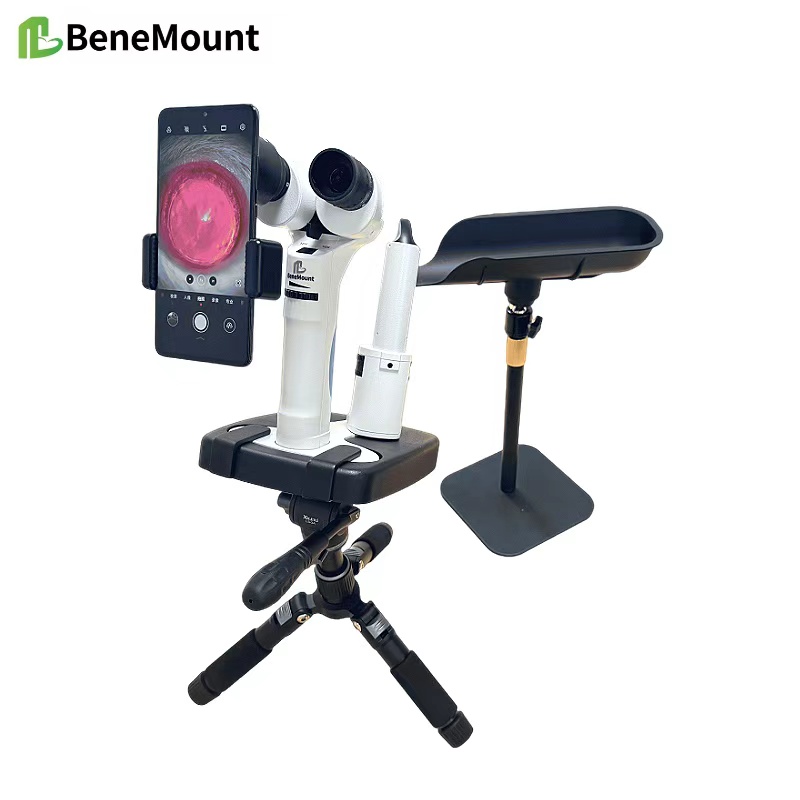
A handheld slit lamp gives the best balance between mobility and precision for mice. The examiner adjusts illumination to a low level so that glare and photophobia do not interfere with imaging. In addition, magnification between 10× and 16× offers enough detail to study corneal and lens structures. The device should remain on a stable surface to reduce hand tremor. Therefore, before every session, the examiner checks alignment and cleans the optics to guarantee reliable performance.
Animal Preparation
Mice need short-acting anesthesia to keep them still during the exam. Isoflurane inhalation is often chosen because recovery is quick and smooth. The animal rests on a warmed platform, which prevents hypothermia. In addition, the examiner positions the mouse carefully to avoid pressure on the neck or chest. This step protects normal circulation and prevents artifacts in the ocular image. To protect the cornea during longer procedures, a drop of lubricant is applied at the beginning of the exam.
Examination Technique
The narrow slit beam allows clear visualization of corneal layers. For example, stromal thinning and epithelial loss become evident under sectioned light. Diffuse illumination provides a broad view of the anterior segment, revealing edema or vascular changes. Retroillumination, on the other hand, highlights lens opacities and posterior corneal deposits by reflecting light from the fundus. As the examiner adjusts the beam width and angle, different structures come into focus. Consequently, ulcers, keratitis, anterior uveitis, and early cataracts can all be documented with accuracy.
Research and Clinical Applications
Slit-lamp imaging of mice supports a wide range of research models. For example, it helps evaluate dry eye, experimental uveitis, and inherited cataracts. Moreover, repeated imaging allows researchers to track disease progression and measure therapeutic response. In pharmacology or gene therapy studies, the slit lamp becomes an objective tool that links structural changes with functional outcomes. Therefore, this method not only enriches research but also enhances the translational value of animal models.
Conclusion
Reliable slit-lamp use requires suitable equipment, careful anesthesia, and skilled illumination control. When researchers follow these principles, they obtain stable and reproducible results. Ultimately, the clinical and scientific significance of slit lamp in mice lies in its ability to reveal subtle ocular changes and to support discoveries that improve both veterinary and human eye health.


