
Canine and Feline Ocular Surface: Essential Guide to Eyelid and Conjunctival Structures
Canine and Feline Ocular Surface
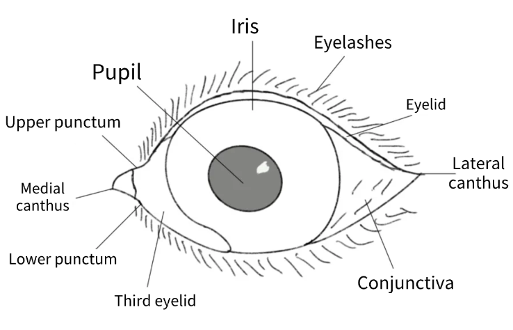
External Eyelid Architecture
The external eyelid system serves as the primary protective barrier between the eye and environmental hazards.
A delicate yet durable layer of hair-bearing skin interfaces with densely arranged cilia that function as microscopic debris collectors.
Below this surface, the orbicularis oculi muscle orchestrates the blink mechanism, distributing tear film across the cornea in under one-third of a second while simultaneously clearing away particulate matter.
The tarsal plate provides structural integrity to the eyelid, securing the palpebral conjunctiva and serving as a foundation for vascular and neural networks.
Any compromise to these structures—whether from injury, cicatricial changes, or neuromuscular dysfunction—undermines blink effectiveness, hastens tear film destabilization, and leaves the cornea vulnerable to dehydration and ulcerative damage.
Meibomian Gland Complex
Within the tarsal plate, approximately 20 to 40 holocrine Meibomian glands are arranged in vertical columns.
These specialized structures discharge lipid-enriched secretions directly onto the eyelid margin, establishing the outermost lipid layer of the tear film.
This sophisticated micro-emulsion reduces aqueous evaporation rates by up to tenfold, creates a smooth optical interface, and generates an antimicrobial barrier concentrated with wax esters and free fatty acids.
Glandular dysfunction—whether obstructive in nature (such as chalazion formation), infectious (meibomianitis), or neoplastic (adenomatous changes)—undermines tear film integrity and triggers compensatory excessive tearing or evaporative dry eye conditions.
Regular magnified examination of Meibomian orifices combined with warm compress therapy for thickened secretions represents an effective preventive approach against chronic ocular surface disorders.
Third Eyelid Structure and Function
The third eyelid consists of a T-shaped cartilaginous framework enveloped by conjunctival tissue, articulating at the medial canthal region.
Its associated lacrimal gland produces approximately 35 to 45 percent of baseline tear volume in both dogs and cats.
During each blink cycle, this membrane sweeps in a dorsolateral direction across the corneal surface, eliminating foreign particles while simultaneously bathing the eye in fresh tear secretions.
Lymphoid follicles integrated within this structure provide initial immune surveillance capabilities, earning it the designation of “conjunctival tonsil.”
Glandular prolapse (commonly termed “cherry eye”) or immune-mediated follicular conjunctivitis can significantly impair both mechanical and secretory functions.
Contemporary surgical approaches favor pocket techniques that preserve rather than remove glandular tissue, thereby preventing postoperative dry eye complications.
Conjunctival-Tear Film Interface
The conjunctiva represents a highly vascularized mucous membrane that lines the posterior eyelids (palpebral portion), reflects at the fornix, and extends onto the globe (bulbar portion).
Goblet cells distributed throughout this tissue secrete MUC5AC mucin, which forms the tear film’s anchoring foundation, while resident conjunctival-associated lymphoid tissue (CALT) generates rapid immunoglobulin A responses.
This tissue demonstrates tolerance toward commensal organisms while maintaining reactivity against pathogenic threats, explaining the pronounced hyperemia and chemosis observed during conjunctival inflammation.
Significantly, conjunctival capillaries function as thermal regulators for corneal temperature, and their permeability characteristics facilitate systemic drug delivery pathways.
Chronic inflammatory processes can deplete goblet cell populations, destabilize tear film composition, and predispose the cornea to exposure-related keratopathy.
Common Pathological Conditions
Blepharitis, characterized by eyelid margin inflammation, frequently results from bacterial colonization, allergic responses, or parasitic infestations.
Clinical manifestations include erythema, edema, and discharge production.
Chronic blepharitis compromises tear film stability and may precipitate corneal irritation.
Conjunctivitis encompasses inflammatory processes characterized by conjunctival erythema, swelling, and discharge.
Etiological factors include bacterial infections, feline herpesvirus, allergic reactions, and keratoconjunctivitis sicca (dry eye syndrome).
Diagnostic evaluation typically incorporates historical assessment, clinical examination, and specific diagnostic procedures such as Schirmer Tear Testing and fluorescein staining to exclude corneal ulceration.
Anatomical abnormalities including entropion (inward eyelid margin rolling) and ectropion (outward eyelid margin rolling) can produce irritation and corneal trauma.
Surgical intervention, such as the Hotz-Celsus procedure, is generally required for correction.
Neoplastic conditions affecting the eyelids, particularly Meibomian gland adenomas in canines and squamous cell carcinomas in felines, demand prompt recognition and therapeutic intervention, typically involving surgical excision.
Diagnostic Protocol
Initial assessment begins with distant observation for blepharospasm, discharge, or periocular swelling, followed by detailed examination of eyelid margins, conjunctival surfaces, and third eyelid structures under focused illumination.
Baseline tear production should be quantified before administering any topical agents using Schirmer Tear Test I; values below 15 mm/min indicate aqueous-deficient dry eye in dogs (felines: below 10 mm/min).
Topical anesthetic should only be instilled after tear testing if intraocular pressure measurement is required—utilizing either rebound or applanation tonometry techniques.
Fluorescein staining should be applied to detect corneal epithelial defects and confirm nasolacrimal duct patency.
In refractory or atypical cases, conjunctival cytology (Diff-Quik staining) and bacterial culture should be obtained; photographic documentation of lesions facilitates monitoring progress.
Canine and Feline Ocular Surface References
- Maggs DJ, Miller PE, Ofri R. Slatter’s Fundamentals of Veterinary Ophthalmology. 4th ed. St Louis: Elsevier; 2008.
- Gelatt KN, Gilger BC, Kern TJ, eds. Veterinary Ophthalmology. 6th ed. Hoboken: Wiley-Blackwell; 2021.
- Merck Veterinary Manual. “Disorders of the Conjunctiva and Eyelids in Small Animals.” Online edition.