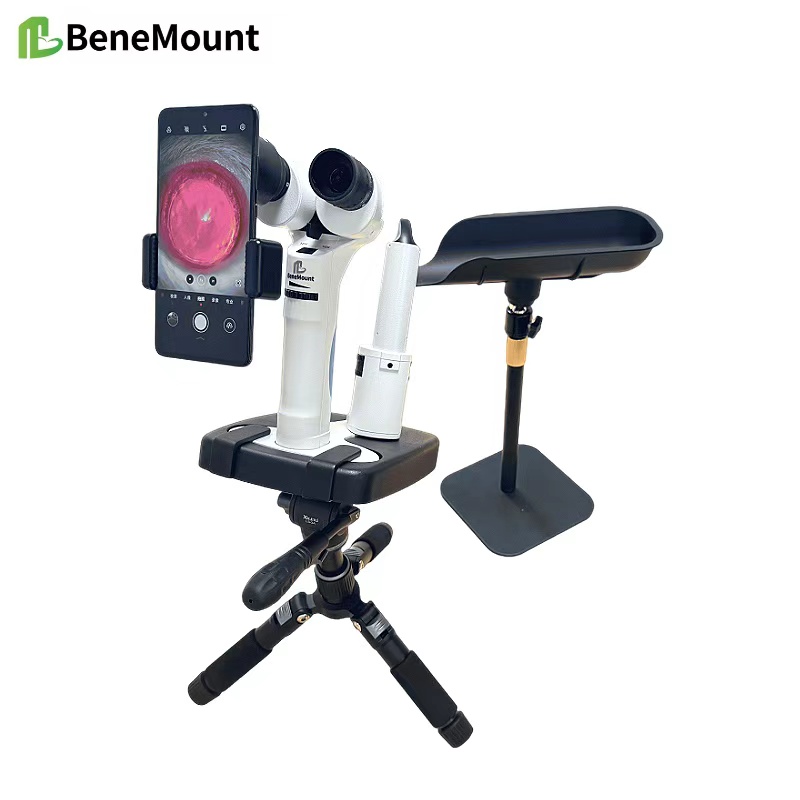
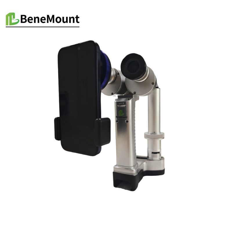
Advanced Slit-Lamp Biomicroscopy expands the diagnostic scope of veterinary ophthalmology by combining the slit lamp with gonioscopy and fundus lenses. This integration allows veterinarians to move beyond anterior segment evaluation and explore deeper ocular structures with accuracy. Such advanced applications bridge the gap between basic biomicroscopy and more complex imaging systems.
Gonioscopy with the Slit Lamp
Gonioscopy involves the use of a specialized lens to visualize the iridocorneal angle. When paired with the slit lamp, it enables detailed inspection of aqueous humor outflow pathways. In dogs predisposed to glaucoma, gonioscopy can reveal pectinate ligament dysplasia or angle narrowing that cannot be seen by routine exam. For cats, angle evaluation assists in detecting secondary glaucoma due to uveitis or lens luxation. By applying a gonioscopy lens under slit-lamp illumination, the clinician can make informed decisions about risk and prognosis.
Fundus Lens Integration
The use of high-quality fundus lenses transforms the slit lamp into an instrument capable of examining the posterior segment. With a condensing lens, the veterinarian gains magnified views of the vitreous, retina, and optic nerve head. Early retinal degeneration, optic nerve pallor, or vitreal opacities become accessible to direct observation. This method is particularly valuable in small animal practice, where subtle posterior abnormalities often require magnification for accurate recognition.
Clinical Advantages
Combining gonioscopy and fundus lenses with slit-lamp biomicroscopy creates a comprehensive approach to ocular assessment. The examiner can evaluate anterior, middle, and posterior structures in a single session. This integration improves efficiency, reduces reliance on more invasive diagnostics, and provides a baseline for monitoring disease progression. For conditions such as glaucoma, uveitis, or retinal degeneration, these advanced applications often determine both treatment strategy and prognosis.
Educational and Research Use
Advanced Slit-Lamp Biomicroscopy is not only a clinical tool but also a valuable method for teaching and research. Veterinary students gain exposure to ocular anatomy beyond the cornea and lens, while researchers employ combined techniques to document disease models with precision. Such versatility reinforces the importance of the slit lamp as a core instrument in both clinical and academic settings.
Conclusion
The integration of gonioscopy and fundus lenses with slit-lamp biomicroscopy extends diagnostic reach far beyond the anterior eye. The true strength of Advanced Slit-Lamp Biomicroscopy lies in its ability to unify multiple techniques into one examination, supporting earlier diagnosis, clearer prognosis, and more effective treatment across species.