Introduction
Knowing how to measure intraocular pressure animals correctly is essential in modern veterinary practice. Eye pressure reflects the delicate balance between the fluid that fills the eye and the outflow channels that drain it. When that balance is disrupted, diseases such as glaucoma or uveitis can quickly threaten vision. For decades, veterinarians struggled with bulky tonometers and procedures that caused stress to patients. Now, portable rebound devices like the iFalcon V100 have made eye-pressure testing simpler, faster, and more comfortable for both animals and clinicians.
Understanding Intraocular Pressure
Intraocular pressure (IOP) is measured in millimeters of mercury (mmHg) and indicates the tension within the eye. Normal ranges vary by species—dogs typically measure between 10–25 mmHg, cats around 15–25 mmHg, and horses 17–28 mmHg. Elevated IOP may signal glaucoma, while unusually low pressure suggests inflammation or trauma.
What makes IOP measurement challenging is that animals cannot be asked to “look straight ahead and don’t blink.” A reliable reading depends on minimizing stress, maintaining the correct head position, and using equipment suited to veterinary patients rather than human eyes.
Choosing the Right Tonometer
There are three main methods for measuring IOP: indentation, applanation, and rebound tonometry. Indentation tonometers, such as the Schiøtz, are largely outdated and require anesthesia. Applanation tonometers flatten the cornea to determine pressure but need numbing drops and precise alignment.
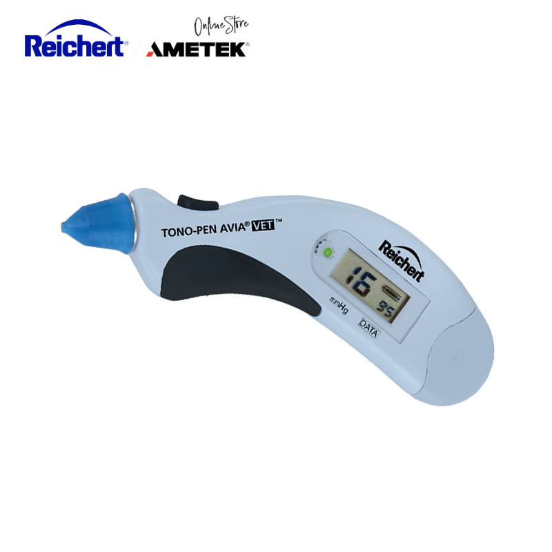
Rebound tonometers, on the other hand, use a light probe that gently touches the cornea and measures the rebound speed. The slower the probe returns, the higher the eye pressure. This method is painless, anesthesia-free, and ideal for small or uncooperative patients.
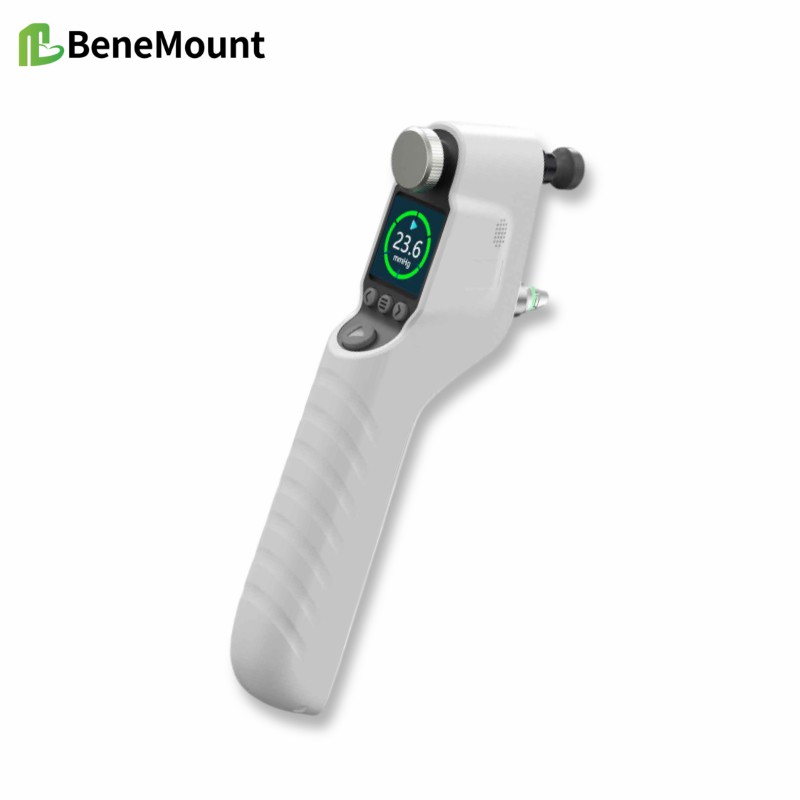
The iFalcon V100 represents the new generation of rebound tonometers designed specifically for animal use. It combines clinical accuracy with true portability, weighing about 220 grams and fitting comfortably in one hand.
Step-by-Step Measurement Technique
- Prepare the Environment
Choose a quiet, dimly lit area to reduce stress. Avoid restraining the animal tightly, as neck or eyelid pressure can artificially raise readings. - Position the Patient
The animal’s eye should be level with the examiner. Dogs and cats can sit or stand naturally; horses may be examined with minimal halter restraint. - Hold the Device Properly
The iFalcon V100 is positioned perpendicular to the cornea at the recommended distance (about 4–8 mm). Its automatic alignment and single-button operation simplify this process. - Take Multiple Readings
The device automatically averages six measurements to improve reliability. The display shows both the mean IOP and a variation index, indicating whether readings are consistent. - Record and Interpret
Document which eye was tested, the species, and any sedation used. Compare values between eyes—differences greater than 8 mmHg often signal a potential problem.
This workflow ensures consistency, whether performed in a clinic, shelter, or field setting.
Why the iFalcon V100 Stands Out
The iFalcon V100 uses rebound technology comparable to leading brands like Icare® but adds practical advantages for everyday veterinary work:
- Rechargeable power for long-term use without disposable batteries
- Large, backlit display that is easy to read even in dim exam rooms
- Memory storage for up to 100 readings to track disease progression
- Species-specific calibration (dog, cat, horse, plus custom mode)
- Self-calibration before each reading for consistent accuracy
Clinicians appreciate its quick setup, reliable results, and the ability to switch between species in seconds. The device’s gentle measurement also means most animals do not require sedation, reducing procedure time and patient stress.
Clinical Applications
Routine IOP measurement is valuable in many situations:
- Glaucoma screening: Detect elevated pressure before permanent vision loss occurs.
- Post-surgical monitoring: Ensure that pressure remains stable after cataract or corneal surgery.
- Uveitis assessment: Confirm low IOP due to inflammation and monitor response to therapy.
- Wellness exams: Include eye pressure checks in senior health plans for early detection of disease.
By integrating tonometry into regular exams, veterinarians improve their diagnostic reach and patient outcomes.
Common Pitfalls and How to Avoid Them
Even with the best instruments, errors can occur. Applying pressure to the eyelid or neck will raise readings, while poor alignment can lead to underestimation. Always ensure the probe is clean, and never reuse disposable tips between patients. If results seem inconsistent, retake measurements after allowing the animal to relax.
Conclusion
Accurate IOP measurement is a cornerstone of animal eye care. With portable rebound devices, the process is now faster, safer, and far more practical. The iFalcon V100 combines comfort for patients with confidence for clinicians, making it an ideal tool for both general and specialty practices. Learning to measure intraocular pressure animals effectively allows veterinarians to detect eye diseases early, monitor progress, and preserve vision across species.