
Animal eye anatomy discover . Detailed visuals explain the cornea, lens, retina, and more for vets and students.
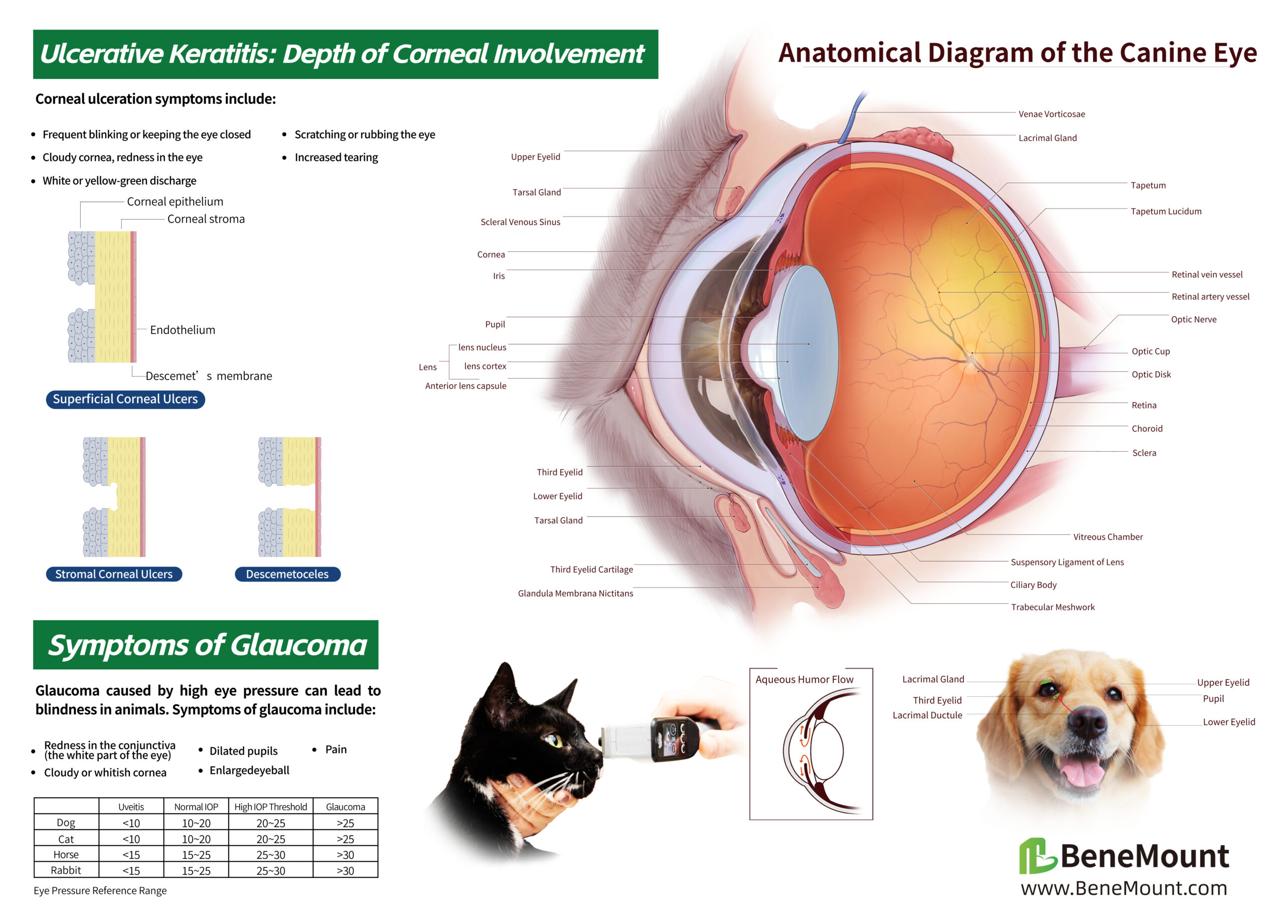
The eye is a crucial visual organ for animals. To understand the various complex ocular diseases in animals, it’s essential to trace them back to their structural origins. Analyzing the anatomy of the animal eye and its surrounding adnexa is very important for studying animal eye diseases and establishing a systematic ophthalmic examination.
Animal Eye Anatomy
An animal’s eye region includes the eyeball, as well as its external structures and adnexa. The basic structure of the eyeball consists of the outermost fibrous tunic (comprising the cornea and sclera), the middle vascular tunic, the innermost neuroectodermal layer (the retina and optic nerve), and the ocular contents (aqueous humor, lens, and vitreous body).
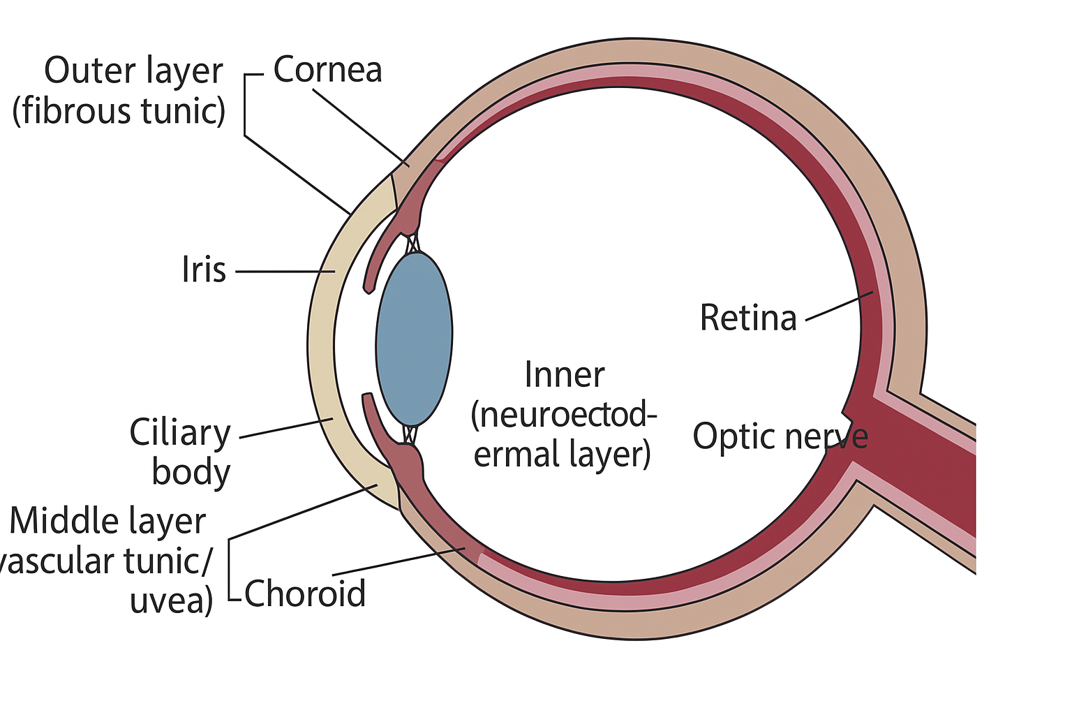
Diagram of the Three-Layered Structure of the Animal Eyeball
Outer layer of the eyeball
1. Cornea
The cornea is the transparent, slightly protruding, disc-shaped thin membrane at the very front of an animal’s eye. It’s thinner in the center and thicker at the edges. It’s completely avascular (lacks blood vessels), though new blood vessels can form on the cornea when it becomes inflamed.
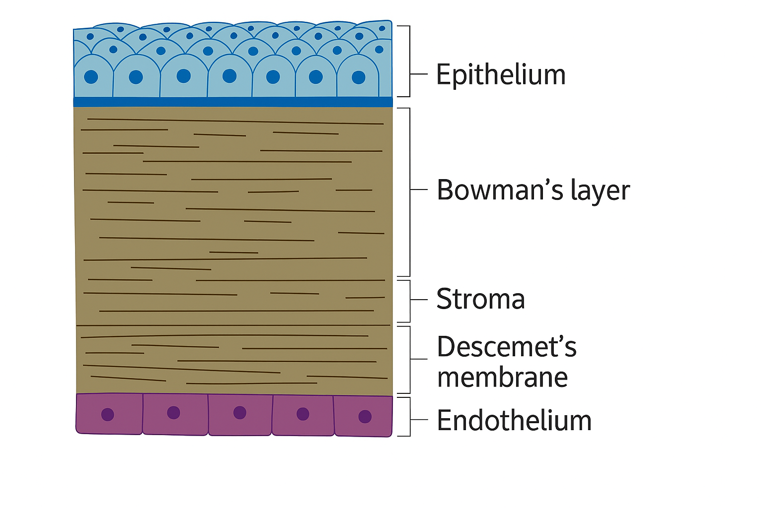
Cross–section of the cornea
The cornea consists of five cell layers, listed from front to back as follows:
- Epithelium
- Basement membrane
- Stroma
- Descemet’s membrane ( also known as lamina elastica posterior)
- Endothelium
2. Sclera
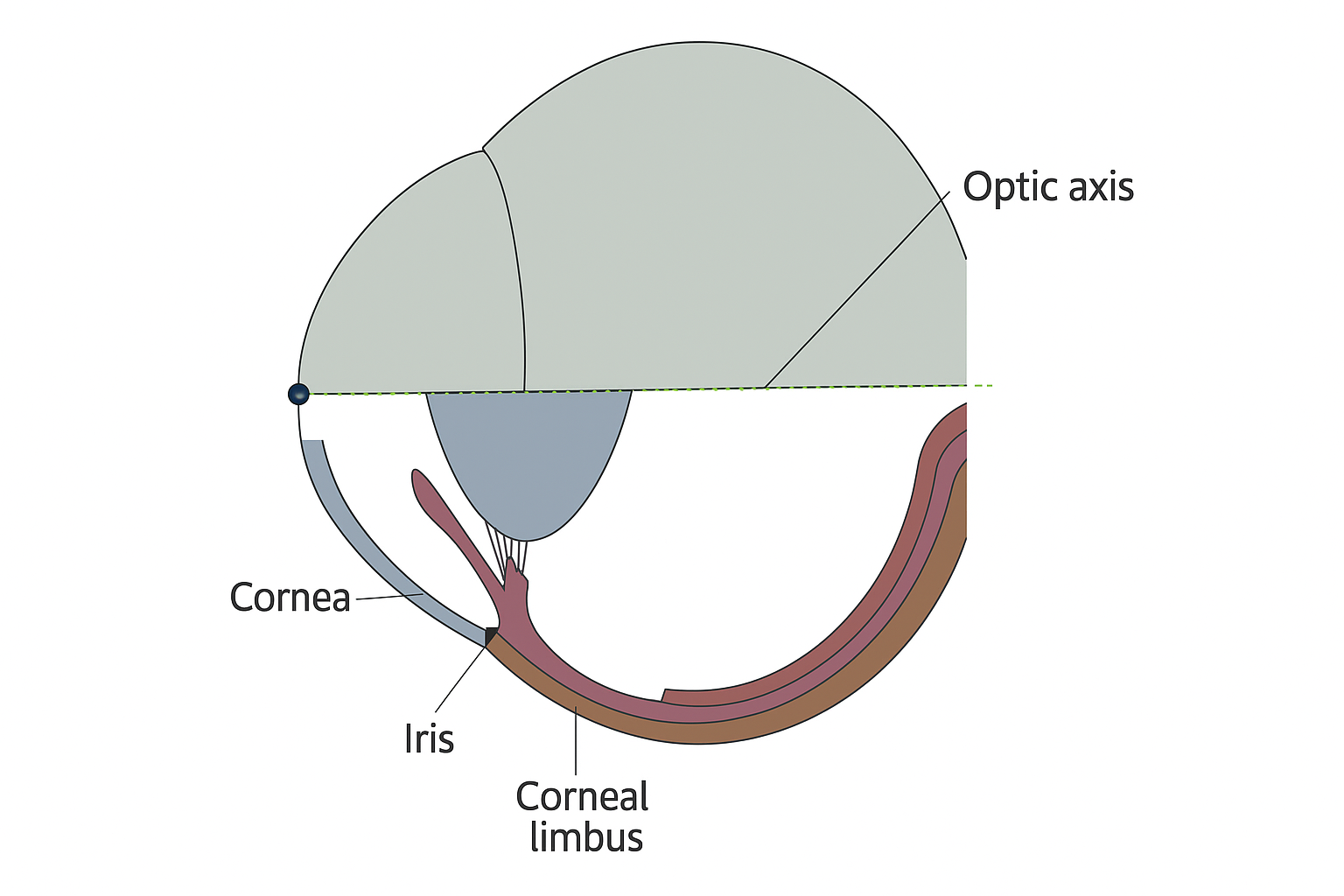
Diagram of the Scleral Sections
The sclera is the outer membrane of the eyeball, a tough and opaque fibrous layer. It’s commonly known as the “white of the eye.”
Middle layer of the eyeball
The middle layer of the eyeball is the vascular layer, also known as the uveal layer. This is a highly vascularized structure, composed of the iris, ciliary body, and choroid.
1. Iris
The iris is the colored part of the eye visible to the naked eye. Its central opening is the pupil. One of the iris’s key functions is to regulate the amount of light entering the eye by changing the size of the pupil. The iris has a rich blood supply, making it prone to intraocular bleeding symptoms if injured.
The color displayed by the iris is caused by variations in the amount of pigment in the iris stroma and epithelium. Brown irises contain more pigment, while blue irises contain less.
2. Ciliarybody

Diagram of the Ciliary Body Sections
The ciliary body is located behind the iris, with a triangular cross-section. Its front connects to the scleral root, and one side connects to the vitreous body. The ciliary body’s functions include producing aqueous humor and regulating the eye’s refractive power through relaxation and contraction.
3. Choroid
The choroid is a dark brown vascular layer situated between the sclera and the retina. It’s rich in blood vessels and pigment granules, providing essential nutrients to the retina.
Inner layer of the eyeball
The inner layer of the eyeball is a nervous membrane, including the retina and the tapetum lucidum.
1. Retina
The retina is a sensory neural tissue and part of the central nervous system. From outer to inner, it is composed of retinal pigment epithelial cells, photoreceptor cells (rods and cones, which are light receptors), bipolar cells, horizontal cells, Müller cells, amacrine cells, and ganglion cells.
From a histological perspective, the layers from outer to inner are: retinal pigment epithelium, photoreceptor layer, outer limiting membrane, outer nuclear layer, outer plexiform layer, inner nuclear layer, inner plexiform layer, ganglion cell layer, nerve fiber layer, and inner limiting membrane.
2. Tapetum Lucidum
Also known as the “choroidal tapetum,” this tissue is present in the eyes of many vertebrates other than humans. Located superior to the optic nerve head, it is a reflective, thin-membrane-like structure within the choroid. This tissue reflects visible light back to the retina, enhancing the retina’s absorption of light and improving the animal’s night vision capabilities.
Ocular Contents

Diagram of Aqueous Humor Circulation
The dynamic balance between the production and drainage of aqueous humor is crucial for maintaining intraocular pressure (IOP). If the production and drainage of aqueous humor become imbalanced, and the fluid cannot drain properly, it will lead to an increase in IOP, thus causing glaucoma. Similarly, if there’s a change in the iridocorneal angle structure or its size, preventing normal aqueous humor drainage, it will also cause an increase in IOP and lead to glaucoma.
2. Lens
The function of the lens is to refract light. It’s composed of transparent protein fibers and is located behind the iris, connected to the ciliary body by the suspensory ligaments. Cataracts occur when the lens proteins denature, causing the lens to become cloudy and opaque.
3. Vitreous Body
The vitreous body is the largest structure within the eyeball, accounting for approximately 80% of its total volume. It’s a transparent, gel-like substance rich in diluted salts, proteins, and hyaluronic acid, and it’s avascular. The primary role of the vitreous body is to help support the retina’s attachment.
Extraocular and Adnexal Structures
1. Orbit
The orbit is the cavity that houses the eyeball. It’s rich in fat, which helps to cushion the eyeball from shock and protect it.
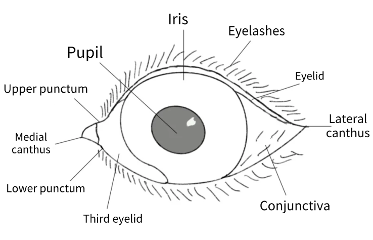
2. Eyelids
Commonly known as “eyelids,” they are divided into upper and lower lids.
They secrete components of the tear film and function to lubricate the eyelids and cornea.Eyelid diseases typically include eyelid inflammation (blepharitis), internal hordeolum (chalazion), and external hordeolum (stye).
3. Eyelashes
Located at the eyelid margin, eyelashes help block foreign objects from entering the eye, preventing damage to the eyeball.
Distichiasis (double rows) or ectopic cilia can rub against the cornea, causing irritating tearing.
4. Tear Film
The tear film protects the cornea and provides it with nutrients.
Abnormal tear film production can lead to dry eye disease. A lack of tear film easily causes corneal damage.
The tear film is primarily composed of three layers: the lipid layer, the aqueous layer, and the mucous layer.
5. Nasolacrimal System
The lacrimal gland, located superolaterally to the eyeball, is responsible for secreting tears.
Tears then enter the superior and inferior puncta near the medial canthus, flow into the lacrimal canaliculi and lacrimal sac, and are finally drained through the nasolacrimal duct into the nasal cavity.
6. Conjunctiva
The conjunctiva is a thin, transparent mucous membrane.
Because it’s rich in blood vessels and highly extensible, it can be used to repair corneal defects.
It also serves as a reservoir for lymphocytes, helping to manage immune responses in the eye. The conjunctiva can be divided into the palpebral conjunctiva and the bulbar conjunctiva.
7. Third Eyelid
Also known as the nictitating membrane, the third eyelid is a movable structure located in the medial canthus.
It’s covered by conjunctival tissue and contains a T-shaped clear cartilage and connective tissue.