Understanding IOP Measurement in Animals
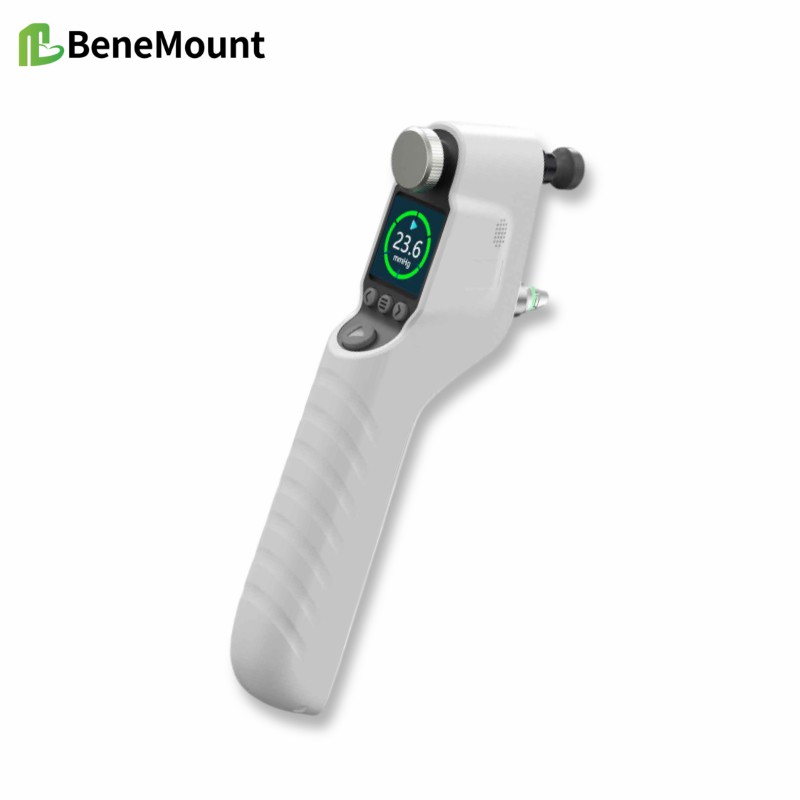
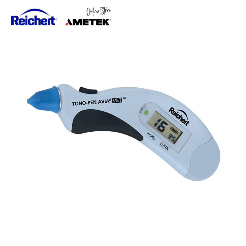
Accurate IOP measurement animals relies not only on the tonometer itself but also on how the patient is positioned and managed during the exam. Posture, stress level, and sedation can all change the reading by several millimeters of mercury. Even with reliable rebound tonometers like the iFalcon V100 or Icare® TONOVET Plus, attention to detail is essential for meaningful data.
Why Technique Matters More Than Equipment
Intraocular pressure reflects the delicate balance between aqueous humor production and drainage. External factors—like restraint, head angle, and lighting—can temporarily disturb that balance. Knowing how to control these variables helps veterinarians separate real ocular change from measurement error.
The Impact of Body Position
How Posture Alters Readings
Changing from standing to lateral recumbency increases venous pressure in the head, often raising eye pressure by 3–6 mmHg. In large dogs and horses, neck compression adds even more. For consistency, test with the head level, eyes at heart height, and spine straight.
Restraint and Handling Technique
Holding the eyelids or neck too firmly can distort venous return. Instead, support the head under the jaw and avoid pressing near the globe. Small adjustments like these make eye pressure readings far more dependable.
Quick Handling Tips
Keep the patient relaxed before testing
Maintain a neutral head angle
Avoid covering or pressing eyelids
Sedation and Its Effect on Eye Pressure
Drugs That Lower or Raise IOP
Different medications influence IOP in different ways. Alpha-2 agonists such as dexmedetomidine lower pressure by reducing fluid production and systemic blood pressure. Agents like ketamine can raise it briefly, while opioids and benzodiazepines have minimal effect. Knowing each drug’s tendency allows clinicians to interpret results correctly.
Timing Matters
Whenever possible, take readings before administering sedatives. If testing must occur under sedation, record the drug type, dose, and time elapsed. This ensures comparisons later are fair and prevents misinterpreting drug effects as disease progression.
Environmental and Equipment Considerations
Creating the Right Testing Environment
Noise, bright lights, and high stress all increase ocular pressure temporarily. A calm, dimly lit room helps animals stay still and reduces the need for restraint. Allow a minute for the patient to settle before testing.
Keeping Devices Reliable
Both the Icare® TONOVET Plus and the iFalcon V100 maintain accuracy when properly calibrated. The iFalcon’s automatic variance monitor flags inconsistent results caused by movement, while the Icare® relies on user awareness. Perform a quick self-check before each session and replace probe tips between patients.
Field Use Tips
Store instruments in padded cases
Let them adjust to room temperature
Avoid humidity and dust near the probe
Building Consistency in Clinical Practice
Standardizing the Process
Every staff member should follow the same approach to positioning and handling. Document posture, sedation status, and device used in every record. This consistency allows small IOP changes to be interpreted as real trends rather than measurement noise.
Why Consistency Improves Detection
When IOP measurement animals follows a routine, fluctuations reveal disease patterns sooner. Stable technique helps detect glaucoma or uveitis earlier, guiding treatment and protecting vision.
Putting It All Together
Accurate IOP measurement animals comes from discipline, not luck. By controlling posture, using gentle restraint, and understanding sedative effects, veterinarians turn simple eye pressure testing into a reliable diagnostic tool. Modern rebound tonometers like the iFalcon V100 and Icare® make that easier—but precision still depends on the hands that hold them.