
Canine Eyelid Tumors: Prevalence and Characteristics Eyelid tumors are the most common type of tumor affecting the canine eye and its adnexa. The majority of these tumors are benign, with reported rates ranging from 73.3% to 87.8%.
Epithelial tissue tumors are far more common than mesenchymal tissue tumors, with a ratio of approximately 5:1, meaning epithelial tumors account for the vast majority. These tumors occur more frequently on the upper eyelid than the lower eyelid. The incidence typically increases in dogs over 10 years of age, with no significant difference observed between sexes.
Patient Information
Here’s the information about the dog:
- Species: Pomeranian
- Age: 10 years old
- Sex: Female
- Weight: 4.4 kg
Chief Complaint
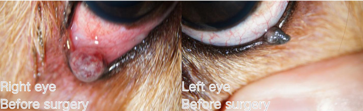
For the past two to three years, the dog has had growths on both lower eyelids. Recently, the growth on the right eye has significantly enlarged, accompanied by redness and swelling of the right conjunctiva, increased eye discharge, and squinting (blepharospasm).
Medical History
The dog has a history of a corneal ulcer in the right eye.
Examination and Diagnosis
Here’s a breakdown of the examinations performed and their findings:
Examination Procedures
- Schirmer Tear Test (STT): This test measures tear production.
- Slit-Lamp Examination: This allows for a magnified view of the eye’s structures.
- Fluorescein Sodium Staining: This dye is used to detect corneal abrasions or ulcers.
Examination Results
- Schirmer Tear Test (STT):
- Right Eye (OD): 25 mm/min
- Left Eye (OS): 17 mm/min
- Slit-Lamp Examination:
- Right Eye (OD): Revealed a significant amount of yellowish-white purulent ocular discharge. The conjunctiva was red and swollen. A red, round mass was observed on the lower eyelid, and the blood vessels within the mass were engorged.
- Left Eye (OS): A dark gray mass was present on the lower eyelid.
- Both Eyes (OU): The cornea appeared normal, and there was no fluorescein sodium staining, indicating no corneal ulcers or abrasions.
Schirmer Tear Test Strips

Fluorescein Sodium Ophthalmic Strips
Diagnosis and Treatment
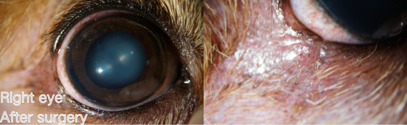
After surgery Right eye
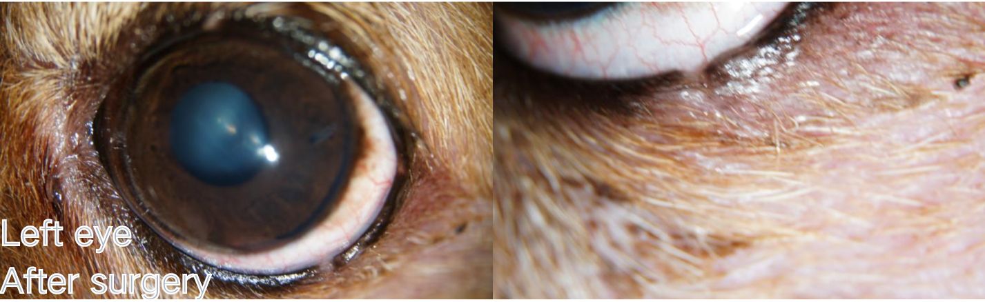
After surgery Left eye
Diagnosis
The diagnosis is bilateral eyelid tumors (OU eyelid tumors) and conjunctivitis in the right eye (OD conjunctivitis).
Treatment Plan
Given the rapid growth of the right eye eyelid tumor, which is rubbing against the eyeball and causing conjunctival redness, inflammation, and a large amount of yellowish-white purulent discharge, surgical excision of the eyelid tumor is recommended to remove the underlying cause. Post-surgery, medication will be administered to treat the conjunctivitis.
Treatment Outcome
The surgical procedure involved a “V”-shaped full-thickness excision of the eyelid tumors from both eyes. The eyelid margins were meticulously closed with 6-0 ophthalmic non-absorbable sutures using a figure-eight pattern, resulting in a smooth and aesthetically pleasing alignment. Following medication, the conjunctivitis resolved.
SMI Ophthalmic Sutures Non-Absorbable
Veterinary Instructions:
Post-surgery, hospitalization is not required. Go home, wear an Elizabethan collar for 24 hours.
Use anti-inflammatory eye drops and systemic anti-inflammatory medication as scheduled. Suture removal in 7-10 days.
Prognosis
The recurrence rate is very low after full-thickness excision of eyelid tumors. We recommended that the owner submit the excised tumor tissue to a laboratory for histopathological examination to determine if it was benign or malignant. If it were malignant, the recurrence rate would be high. In this case, the owner declined the submission.
Summary of Canine Eyelid Tumors
Eyelid tumors are common in older dogs. They are mostly meibomian adenomas. Most are benign, at least 75%. Few are malignant, like melanoma or fibrosarcoma. Cats, however, often have malignant eyelid tumors.
Surgery is advised for irritation. This includes conjunctivitis, ulcers, or rapid growth. Observe or use drops if symptoms are mild.
Other methods, like cryotherapy, have high recurrence. “V” or “wedge” excisions are best. Direct closure works if excision is small. Larger excisions need eyelid reconstruction. Use 6-0 non-absorbable sutures. Keep sutures off the conjunctiva. Tools like eyelid plates can help.
Local anesthesia is often possible. This avoids general anesthesia risks. A 1mm margin is standard. Always test tumors for malignancy. Malignant tumors need wider excision and more treatment.