
Understanding Glaucoma in Dogs
Glaucoma is a group of diseases characterized by elevated intraocular pressure (IOP). This increased pressure directly compresses the optic nerve, leading to various symptoms and potentially blindness. Clinically, if a dog’s IOP is above 25 mmHg, glaucoma is suspected [1-2].
Canine glaucoma is incurable. Managing IOP with eye drops and surgery becomes increasingly difficult over time, ultimately leading to blindness. However, when there’s persistent ocular pain and discomfort, or severe corneal epithelial damage develops due to conditions like exposure keratitis, surgical intervention becomes necessary for a blind, glaucomatous eye. The primary goals of such surgery are to alleviate glaucoma-induced eye pain and discomfort and to terminate the glaucoma itself [3].
Case Report: Prosthetic Eye After Corneal Endothelial Rupture Caused by Glaucoma
This paper presents the case of a dog with glaucoma. After being diagnosed at an external clinic, surgical intervention was not performed. The glaucoma subsequently led to corneal endothelial rupture. The dog was then referred to our hospital for bilateral prosthetic eye implantation. Both corneas and prosthetic eyes recovered well, with no adverse outcomes. The diagnostic and treatment process is detailed below for your reference.
Patient Information
Species: Canine
Age: 9 years old
Sex: Female
Weight: 10 kg
Chief Complaint: The owner noticed the dog bumping into walls at home and observed abnormalities in her eyeballs. After an unproductive visit to a veterinary clinic, they opted for conservative management at home.
History: Diagnosed with glaucoma two months prior at an external clinic. No surgical intervention was performed.
Clinical Examination
Vital Signs:
Temperature: 39.2℃
Heart Rate: 140 bpm
Respiration Rate: 50 breaths/min
Body Condition Score (BCS): 6/9
Ophthalmic Examination
| Examination Item | OD (Right Eye) | OS (Left Eye) |
|---|---|---|
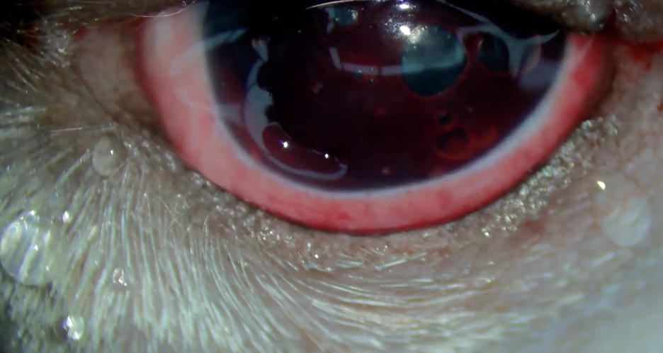
| Intraocular Pressure | 42 | 53 |
| STT | 15 | 15 |
| Fluorescein Staining | No obvious staining | Obvious corneal epithelial staining, no corneal uptake |
| Slit Lamp | Lens dislocation into anterior chamber | Corneal epithelial staining observed |
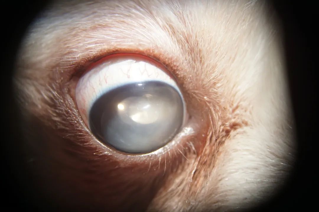
OD Lens dislocation into anterior chamber
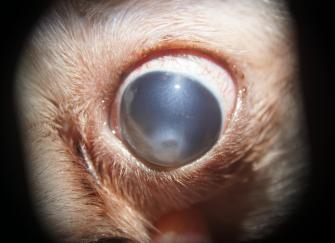
OS Corneal epithelial staining observed
Diagnosis and Suspected Directions
Unfortunately, the external clinic’s examination results for this case were not preserved. Upon presentation to our hospital, the patient was found to be completely blind. Furthermore, the ideal window for a glaucoma drainage valve procedure had been missed.
Treatment and Outcome
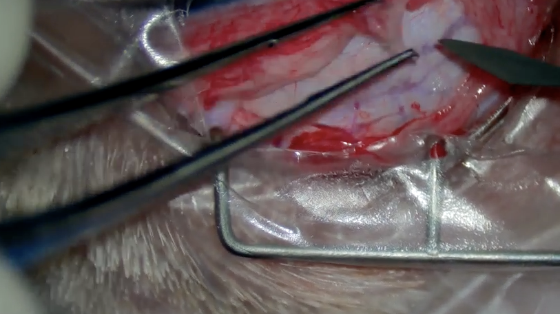
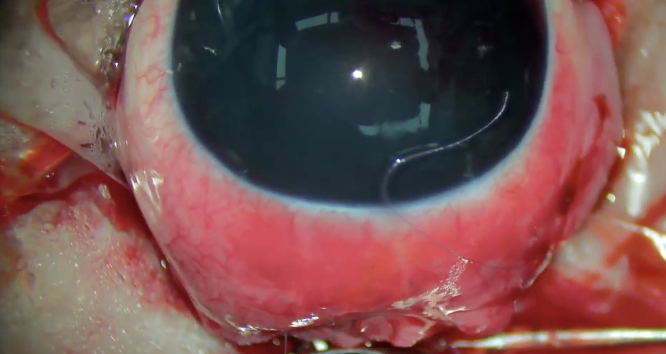
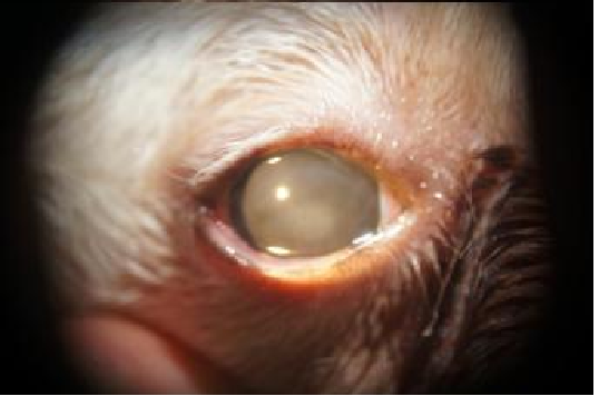
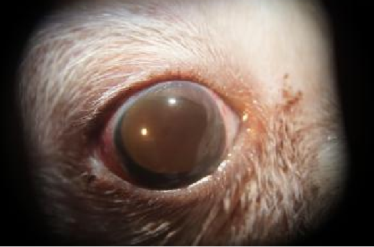
Surgical Treatment: Bilateral prosthetic eye surgery was performed.
(Note: In this case, the left eye had extremely high intraocular pressure, leading to an endothelial ulcer.)
Glaucoma in Dogs
Treatment Protocol
Intravenous Therapy: Anti-inflammatory and pain relief medication administered for one week.
Oral Medication: Health supplement, two anthocyanin capsules given twice daily (morning and evening).
Prognosis
4-6 weeks post-surgery: The cornea recovered well, and the prosthetic eye recovered well.
Summary
- Examine Unaffected Eye: Due to the bilateral nature of primary glaucoma, veterinarians must examine the unaffected eye to determine its risk of developing glaucoma.
- Prophylactic Treatment for Susceptible Eyes: For susceptible, unaffected eyes at risk of glaucoma, consider prophylactic treatment such as 0.25% demecarium bromide (miotic) eye drops every 12 hours, or 0.5% timolol maleate every 12 hours, or 2% dorzolamide every 8-12 hours.
- Possible Complications: Potential complications include blindness and chronic ocular pain.
- Long-Term Medical Management for Chronic Disease: Glaucoma is a chronic condition that requires long-term medical treatment, even after surgical intervention.
- Medication Alone Often Leads to Blindness: Most cases treated solely with medication will eventually lead to blindness. (If blindness occurs, consider prosthetic eye surgery).
- Glaucoma Drainage Valve Offers Better Vision Preservation: Surgical treatment with a glaucoma drainage valve offers a better chance of long-term vision preservation.
- Glaucoma Secondary to Lens Luxation: The prognosis may be good if the lens is successfully removed and followed by appropriate postoperative medical treatment.
- Glaucoma Secondary to Anterior Uveitis: The prognosis may be good once the uveitis is controlled.