
Veterinary Ophthalmology Case Reference: A 7-Year-Old Dog’s Sudden Blindness
This report details the case of a 7-year-old mixed-breed dog that experienced sudden onset blindness. Initially, routine ophthalmic examinations at an external clinic revealed no obvious external eye abnormalities, leading to a referral to our facility.
Comprehensive systemic imaging (CT, MRI), along with hematological and neurological assessments, ultimately revealed a nasal/sinus mass that had invaded the olfactory bulb intracranially, causing the sudden blindness.
This case highlights how a superficial symptom like “blindness” can lead to the discovery of a deeper, intracranial lesion. It emphasizes the critical importance of a complete visual pathway examination and multidisciplinary collaboration in such diagnoses.
The following outlines the diagnostic and treatment process for this case, offered for your reference:
Patient Information
Species: Canine
Age: 7 years old
Sex: Female
Breed: Mixed-breed
Chief Complaint: Sudden onset blindness on November 30, 2024. No abnormalities found during initial local veterinary visit, leading to referral to our hospital.
History: Discomfort began on November 17, with neck pain and subsequent vision decline. On November 28, she experienced epistaxis (nosebleed) from the right side. Sneezing had been occurring for 2-3 months. On November 29, the owner noticed a significant decline in vision, and by November 30, she was completely blind and had experienced considerable epistaxis again.
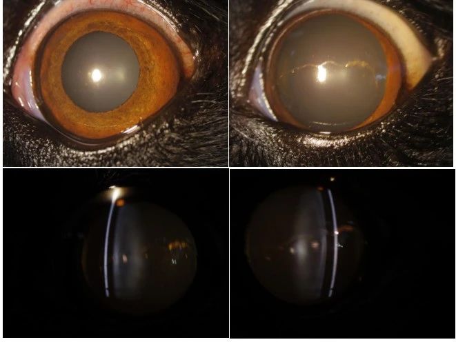
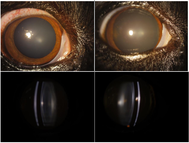
Physical Exam Findings: Both eyes appeared grossly normal on visual inspection. In the examination room, the dog would bump into furniture, indicating impaired vision.
Examinations
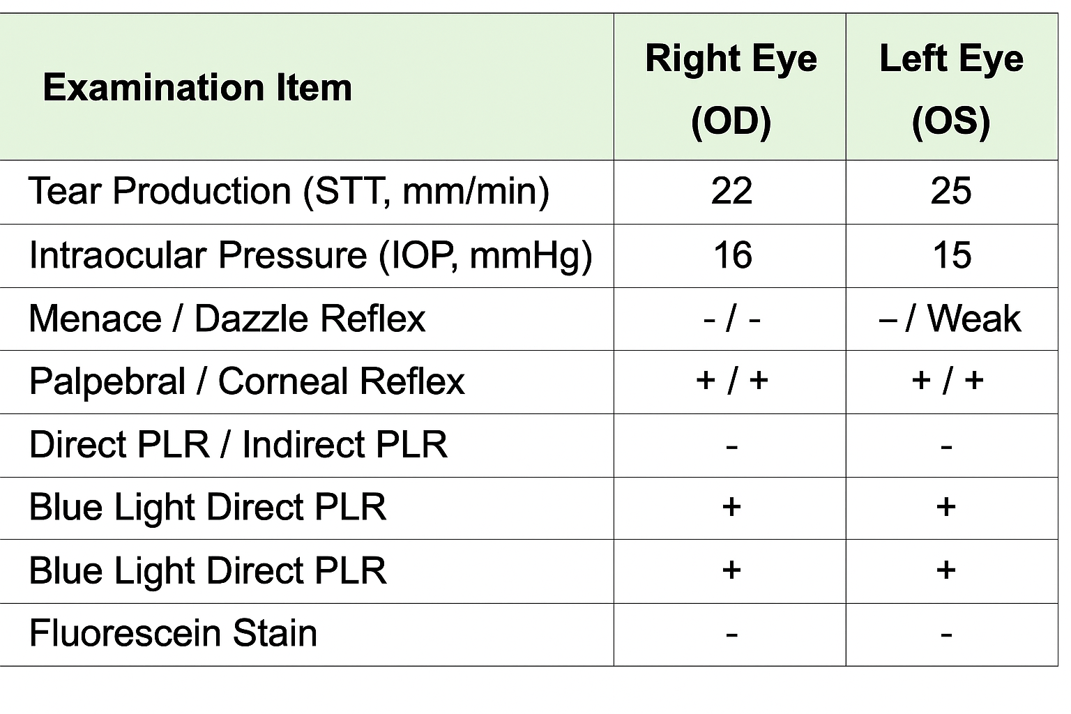
Ophthalmic Examination: Abnormalities were noted in the visual examination.
Neurological Examination: No obvious abnormalities were found.
Veterinary Ophthalmology Case Reference
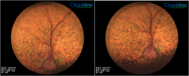
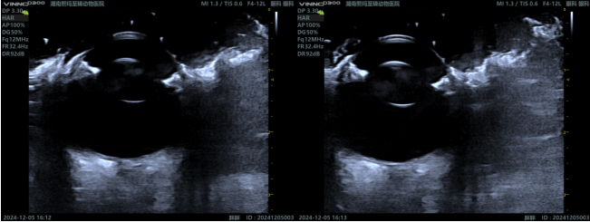
Left Eye Examination
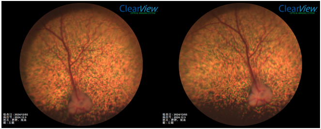
Fundus Photography:
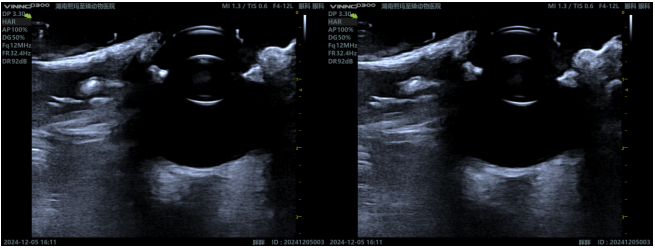
Ocular Ultrasound:
Right Eye Examination
Fundus Photography:
Ocular Ultrasound:
Ancillary Diagnostics
Abdominal Ultrasound & DR (Digital Radiography): No significant abnormalities were observed.
Bloodwork: Revealed an inflammatory response, along with elevated alkaline phosphatase and alanine transaminase level.
CT and MRI Examinations:
Imaging Description:
MRI: A significant lesion was identified in the right olfactory bulb.
- T2WI sequence: Hyperintense (bright signal)
- T1WI sequence: Hypointense (dark signal)
- FLAIR sequence: Hyperintense (bright signal)
- Post-contrast: No significant enhancement was observed.
CT: There were prominent proliferative or mass-like lesions in the nasal cavity, paranasal sinuses, and nasopharynx, accompanied by ethmoid bone lysis. The lesion in the right nasal cavity was more severe than in the left.
- The cribriform plate structure between the nasal cavity and the telencephalon was incomplete, showing bone destruction.
- Nasal cavity contents showed heterogeneous enhancement after contrast administration.
- An irregularly enhancing area (approximately 12.4 x 12.5 mm) was present in the right telencephalic olfactory bulb after contrast administration
Imaging Impressions:
- Nasal mass / Sinusitis / Nasal polyp (nature to be determined)
- Brain tumor / Encephalitis (nature to be determined)
Diagnosis
Preliminary Diagnosis
- Based on the clinical signs (sudden vision loss, epistaxis, etc.) and imaging findings, we highly suspect a nasal cavity/sinus tumor that has invaded the cranium, affecting the visual center (specifically the olfactory bulb and cribriform plate area at the front of the visual pathway), leading to acute blindness.
- The possibility of localized ocular disease (such as SARDS, PRA, optic neuritis) has been temporarily ruled out by routine ophthalmic examinations.
- While nuclear sclerosis is present, it’s not sufficient to explain the sudden blindness and neuroimaging changes.
Key Differential Diagnoses for Blindness
- Ocular globe lesions: Cataracts, corneal diseases, glaucoma, uveitis, retinal degeneration, etc. Most of these have been ruled out by routine eye exams in this case.
- Optic nerve lesions: Inflammation (optic neuritis), ischemia, tumors, etc. Fundus examination showed no obvious optic disc abnormalities, but MRI has already indicated an intracranial lesion.
- Visual pathway/central nervous system lesions: Brain tumors, hemorrhage, inflammation, or infarction, etc. The imaging changes in this case support a central nervous system lesion.
Treatment Approach and Plan
Further Clarification of Pathological Nature
- Biopsy: The nasal/nasopharyngeal mass and intracranial changes should undergo a biopsy (either via rhinoscopic biopsy or surgical exploration) to confirm whether it’s a malignant tumor (such as nasal adenocarcinoma, osteosarcoma, fibrosarcoma, lymphoma, etc.) or an inflammatory lesion.
- Treatment Suitability: If confirmed to be a malignant tumor, we’ll need to assess its suitability for surgical resection, radiation therapy, or chemotherapy.
Symptomatic and Supportive Treatment
- Prevent Complications: Depending on the situation, we may use hemostatic drugs (for persistent bleeding), dehydrating agents, or corticosteroids (if inflammation or edema is significant; however, hormone use in tumor patients requires careful consideration).
- Pain Management: For neck and back pain, analgesics can be administered, and the patient should be closely monitored.
Prognosis
- Outlook: If confirmed to be an intracranial tumor or a severe nasal tumor invading the orbit and brain structures, the prognosis is often guarded to poor.
- Vision Recovery: The likelihood of vision recovery is low. We’ll need to discuss with the owner that the treatment goals will likely focus more on pain relief, bleeding control, and maintaining quality of life.
- Extended Survival: If diagnosed promptly and effective surgical, chemotherapy, or radiation therapy can be performed, some cases may see an extended survival time and improved clinical symptoms.
Case Revelations and Summary
- Consider the Entire Visual Pathway for Vision Impairment: When an animal experiences vision loss or blindness, don’t limit the investigation to just ophthalmic diseases. It’s crucial to also consider the central components of the visual pathway, including the optic nerve, optic chiasm, optic tracts, lateral geniculate body, optic radiations, and visual cortex.
- Co-occurrence of Respiratory and Neurological Symptoms: If upper respiratory symptoms like epistaxis (nosebleeds) and sneezing are present alongside neurological signs, be wary of potential spread or invasion of nasal cavity or sinus lesions to surrounding structures, especially brain tissue.
- Importance of Imaging for Diagnosis: CT and MRI scans are essential for assessing tumor staging, extent, and the degree of damage to surrounding tissues.
- Multidisciplinary Collaboration: When ophthalmic examinations can’t explain the cause, particularly when intracranial lesions are involved, it’s necessary to collaborate with teams from neurology, imaging, and other departments to formulate a comprehensive diagnosis and treatment plan.